

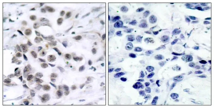
IHC-P analysis of human breast carcinoma tissue using GTX50463 Chk1 antibody. Left : Primary antibody Right : Primary antibody pre-incubated with the antigen specific peptide

IHC-P analysis of human breast carcinoma tissue using GTX50463 Chk1 antibody. Left : Primary antibody Right : Primary antibody pre-incubated with the antigen specific peptide
Chk1 antibody
GTX50463
ApplicationsWestern Blot, ImmunoHistoChemistry, ImmunoHistoChemistry Paraffin
Product group Antibodies
ReactivityHuman
TargetCHEK1
Overview
- SupplierGeneTex
- Product NameChk1 antibody
- Delivery Days Customer9
- Application Supplier NoteWB: 1:500-1:1000. IHC-P: 1:50-1:100. *Optimal dilutions/concentrations should be determined by the researcher.Not tested in other applications.
- ApplicationsWestern Blot, ImmunoHistoChemistry, ImmunoHistoChemistry Paraffin
- CertificationResearch Use Only
- ClonalityPolyclonal
- Concentration1 mg/ml
- ConjugateUnconjugated
- Gene ID1111
- Target nameCHEK1
- Target descriptioncheckpoint kinase 1
- Target synonymsCHK1, OZEMA21, serine/threonine-protein kinase Chk1, CHK1 checkpoint homolog, Checkpoint, S. pombe, homolog of, 1, Chk1-S, cell cycle checkpoint kinase
- HostRabbit
- IsotypeIgG
- Protein IDO14757
- Protein NameSerine/threonine-protein kinase Chk1
- Scientific DescriptionThe protein encoded by this gene belongs to the Ser/Thr protein kinase family. It is required for checkpoint mediated cell cycle arrest in response to DNA damage or the presence of unreplicated DNA. This protein acts to integrate signals from ATM and ATR, two cell cycle proteins involved in DNA damage responses, that also associate with chromatin in meiotic prophase I. Phosphorylation of CDC25A protein phosphatase by this protein is required for cells to delay cell cycle progression in response to double-strand DNA breaks. Several alternatively spliced transcript variants have been found for this gene. [provided by RefSeq, Oct 2011]
- ReactivityHuman
- Storage Instruction-20°C or -80°C,2°C to 8°C
- UNSPSC12352203
References
- Read ML, Fong JC, Modasia B, et al. Elevated PTTG and PBF predicts poor patient outcome and modulates DNA damage response genes in thyroid cancer. Oncogene. 2017,36(37):5296-5308. doi: 10.1038/onc.2017.154Read this paper
Datasheet
Related products

Product group Antibodies
Anti-Chk1/CHEK1 Antibody Picoband(r)A01060-CARRIER-FREE
ApplicationsFlow Cytometry, Western Blot
ReactivityHuman
TargetCHEK1
- SizePrice
Product group Antibodies
Chek1 Recombinant AntibodyCAC12088
ApplicationsImmunoFluorescence, Western Blot, ELISA, ImmunoHistoChemistry
TargetCHEK1
- SizePrice
Product group Antibodies
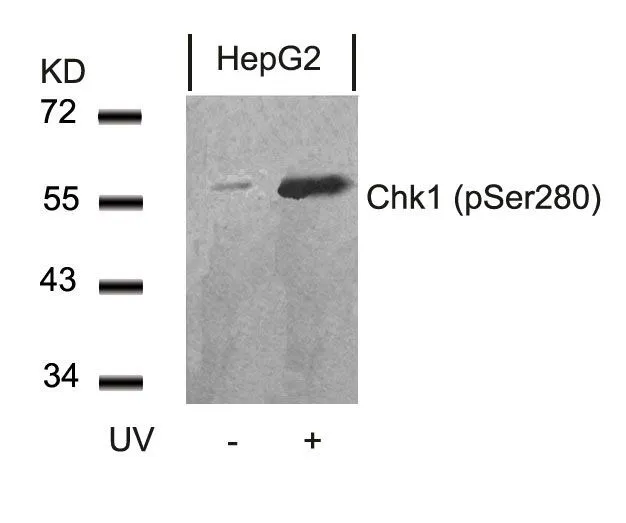
Chk1 (phospho Ser280) antibodyGTX50200
ApplicationsWestern Blot
ReactivityHuman
TargetCHEK1
- SizePrice
Product group Antibodies
Chk1 antibody [6F5]GTX70303
ApplicationsImmunoFluorescence, Western Blot, ImmunoCytoChemistry
ReactivityHuman
TargetCHEK1
- SizePrice
Product group Antibodies
Chk1 (phospho Ser345) antibodyGTX79111
ApplicationsImmunoHistoChemistry, ImmunoHistoChemistry Paraffin
ReactivityHuman
TargetCHEK1
- SizePrice
![WB analysis of HEK293T cells transfected with the pCMV6-ENTRY control (1) and pCMV6-ENTRY CHK1 cDNA (2) using GTX83385 Chk1 antibody [2G1D5].](https://www.genetex.com/upload/website/prouct_img/normal/GTX83385/GTX83385_20170912_WB_1_w_23061322_728.webp)
Product group Antibodies
References
Chk1 antibody [2G1D5]GTX83385
ApplicationsImmunoFluorescence, Western Blot, ELISA, ImmunoCytoChemistry
ReactivityHuman, Mouse
TargetCHEK1
- SizePrice
Product group Antibodies
Anti-CHEK1 Antibody144-07653
ApplicationsImmunoFluorescence, Western Blot, ImmunoHistoChemistry
ReactivityHuman, Mouse, Rat
TargetCHEK1
- SizePrice
Product group Antibodies
Chk1 (phospho Ser296) antibodyGTX55053
ApplicationsWestern Blot, ImmunoHistoChemistry, ImmunoHistoChemistry Paraffin
ReactivityHuman
TargetCHEK1
- SizePrice
Product group Antibodies
Chk1 antibodyGTX56248
ApplicationsImmunoFluorescence, Western Blot, ImmunoCytoChemistry, ImmunoHistoChemistry, ImmunoHistoChemistry Paraffin
ReactivityHuman, Mouse, Rat
TargetCHEK1
- SizePrice