

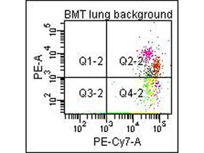
Flow Cytometry of Anti-Collagen Type I Biotin Conjugated Antibody. Cells: mouse lung. Stimulation: none. Primary antibody: biotin conjugated anti-collagen type I antibody. Secondary antibody: PE-conjugated CD45 and PE-conjugated anti-collagen type I secondary antibody.
. Lane 1: Wistar rat hepatic stellate cells (HSC) in control (GFP-transduced). Lane 2: PPARg-transduced cell lysates. Load: 100 μg per lane. Protein staining shown below each blot depicts equal protein loading. Primary antibody: anti-Collagen I antibody at 0.2–2 μg/10 ml for overnight at 4oC. Secondary antibody: horseradish peroxidase-conjugated rabbit secondary antibody at 1 μg/10 ml for overnight at 4oC. Block: TBS with 5% Non-fat milk. Predicted/Observed size: 138.9 kDa for Collagen I. Other band(s): none.")
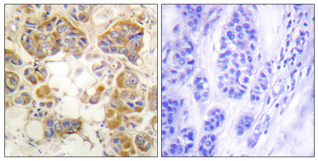
. Tissue: Human Skin at pH9. Fixation: formalin fixed paraffin embedded. Antigen retrieval: not required. Primary antibody: Collagen Type I antibody at 10 μg/mL for 1 h at RT. Secondary antibody: Peroxidase rabbit secondary antibody at 1:10,000 for 45 min at RT. Localization: Collagen Type I is secreted in the extracellular matrix. Staining: Collagen Type I as precipitated brown signal (A) with hematoxylin purple nuclear counterstain. With corresponding negative control (B)")
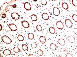
. Tissue: Human Skin at pH6. Fixation: formalin fixed paraffin embedded. Antigen retrieval: not required. Primary antibody: Collagen Type I antibody at 10 μg/mL for 1 h at RT. Secondary antibody: Peroxidase rabbit secondary antibody at 1:10,000 for 45 min at RT. Localization: Collagen Type I is secreted in the extracellular matrix. Staining: Collagen Type I as precipitated brown signal (A) with hematoxylin purple nuclear counterstain. With corresponding negative conrol (B)")

. Panel A : Positive control Panel B : Negative control Dilution : 10 μg/mL")
. Panel A : Positive control Panel B : Negative control Dilution : 10 μgm/mL")
. Left lane : GFP-transduced Right lane : PPARg-transduced Loading : 100 μg")
Flow Cytometry of Anti-Collagen Type I Biotin Conjugated Antibody. Cells: mouse lung. Stimulation: none. Primary antibody: biotin conjugated anti-collagen type I antibody. Secondary antibody: PE-conjugated CD45 and PE-conjugated anti-collagen type I secondary antibody.
Collagen I antibody (Biotin)
GTX26577
ApplicationsDot Blot, Flow Cytometry, ImmunoFluorescence, Western Blot, ImmunoCytoChemistry, ImmunoHistoChemistry, ImmunoHistoChemistry Paraffin, Other Application
Product group Antibodies
ReactivityBovine, Human, Mouse, Rat
TargetCOL1A1
Overview
- SupplierGeneTex
- Product NameCollagen I antibody (Biotin)
- Delivery Days Customer9
- Application Supplier NoteWB: 1:3000-1:6000. IHC-P: 1:50-1:200. *Optimal dilutions/concentrations should be determined by the researcher.Not tested in other applications.
- ApplicationsDot Blot, Flow Cytometry, ImmunoFluorescence, Western Blot, ImmunoCytoChemistry, ImmunoHistoChemistry, ImmunoHistoChemistry Paraffin, Other Application
- CertificationResearch Use Only
- ClonalityPolyclonal
- Concentration1 mg/ml
- ConjugateBiotin
- Gene ID1277
- Target nameCOL1A1
- Target descriptioncollagen type I alpha 1 chain
- Target synonymsCAFYD, EDSARTH1, EDSC, OI1, OI2, OI3, OI4, collagen alpha-1(I) chain, alpha-1 type I collagen, alpha1(I) procollagen, collagen Col1-ColIII-1, collagen Col1-ColIII-2, collagen alpha 1 chain type I, collagen alpha-1(I) chain preproprotein, collagen of skin, tendon and bone, alpha-1 chain, collagen, type I, alpha 1, pro-alpha-1 collagen type 1, type I proalpha 1, type I procollagen alpha 1 chain
- HostRabbit
- IsotypeIgG
- Protein IDP02452
- Protein NameCollagen alpha-1(I) chain
- Scientific DescriptionThis antibody is well suited to detect extracellular matrix proteins in normal as well as disease state tissues. Disruption of tissue organization is the hallmark of neoplasia. Malignant lesions can be distinguished from benign by examining the breakdown of basement membranes and loss of 3-dimensional architecture. Malignant cells are presumed to use matrix metalloproteases to degrade barriers created by the extracellular matrix, which then allows metastasis to occur. Collagenases, stomelysins and gelatinases can collectively degrade all of the various components of the extracellular matrix, including fibrillar and non-fibrillar collagens and basement membrane glycoproteins.
- ReactivityBovine, Human, Mouse, Rat
- Storage Instruction-20°C or -80°C,2°C to 8°C
- UNSPSC41116161
Datasheet
Related products
Product group Antibodies
Anti-Collagen I AntibodyA95083
ApplicationsImmunoFluorescence, ELISA, ImmunoHistoChemistry
ReactivityHuman, Mouse, Rat
- SizePrice
Product group Antibodies
Anti-COL1A1 Antibody144-62810
ApplicationsImmunoFluorescence, ImmunoPrecipitation, Western Blot, ImmunoHistoChemistry
ReactivityHuman, Mouse, Rat
TargetCOL1A1
- SizePrice
Product group Antibodies

Anti-collagen type I [3P1-31], Human IgG1-Fc Fusion,AB04222-10.159
ApplicationsELISA
ReactivityHuman
TargetCOL1A1
- SizePrice
Product group Antibodies

COL1A1 / Collagen I Alpha 1 AntibodyLS-C831483
ApplicationsImmunoHistoChemistry
ReactivityHuman
TargetCOL1A1
- SizePrice

Product group Antibodies
References
Collagen I Polyclonal AntibodyBS-10423R
ApplicationsFlow Cytometry, ImmunoFluorescence, Western Blot, ImmunoCytoChemistry, ImmunoHistoChemistry, ImmunoHistoChemistry Frozen, ImmunoHistoChemistry Paraffin
TargetCOL1A1
- SizePrice
Product group Antibodies
COL1A1 Monoclonal AntibodyCSB-MA139659
ApplicationsELISA, ImmunoHistoChemistry
ReactivityHuman, Mouse, Rat
TargetCOL1A1
- SizePrice
Product group Antibodies

Collagen Type I, humanCO20111-0.1
ApplicationsImmunoFluorescence, Western Blot, ELISA, ImmunoHistoChemistry, ImmunoHistoChemistry Paraffin, RadioImmunoAssay
TargetCOL1A1
- SizePrice
Product group Antibodies
ApplicationsFlow Cytometry
TargetCOL1A1
- SizePrice
Product group Antibodies
References
Collagen I antibodyGTX20292
ApplicationsDot Blot, Flow Cytometry, ImmunoFluorescence, ImmunoPrecipitation, Western Blot, ELISA, ImmunoCytoChemistry, ImmunoHistoChemistry, ImmunoHistoChemistry Paraffin, Other Application
ReactivityBovine, Hamster, Human, Mouse, Porcine, Rat
TargetCOL1A1
- SizePrice
Product group Antibodies
References
COL1A1 antibodyGTX82720
ApplicationsImmunoFluorescence, Western Blot, ImmunoCytoChemistry, ImmunoHistoChemistry, ImmunoHistoChemistry Paraffin
ReactivityHuman, Mouse, Rat, Sheep
TargetCOL1A1
- SizePrice