

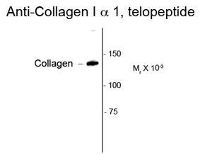
Western blot of rat lung lysate showing specific immunolabeling of the ~140k collagen protein using COL1A1 antibody (GTX82720).
 that has formed fibrils in the extracellular matrix")
Western blot of rat lung lysate showing specific immunolabeling of the ~140k collagen protein using COL1A1 antibody (GTX82720).
COL1A1 antibody
GTX82720
ApplicationsImmunoFluorescence, Western Blot, ImmunoCytoChemistry, ImmunoHistoChemistry, ImmunoHistoChemistry Paraffin
Product group Antibodies
ReactivityHuman, Mouse, Rat, Sheep
TargetCOL1A1
Overview
- SupplierGeneTex
- Product NameCOL1A1 antibody
- Delivery Days Customer9
- Application Supplier NoteWe recommend the following starting dilutions: For WB: Use at a dilution of 1:1000. For IHC: Use at a dilution of 1:100. Optimal dilutions should be determined by the end user.
- ApplicationsImmunoFluorescence, Western Blot, ImmunoCytoChemistry, ImmunoHistoChemistry, ImmunoHistoChemistry Paraffin
- CertificationResearch Use Only
- ClonalityPolyclonal
- ConjugateUnconjugated
- Gene ID1277
- Target nameCOL1A1
- Target descriptioncollagen type I alpha 1 chain
- Target synonymsCAFYD, EDSARTH1, EDSC, OI1, OI2, OI3, OI4, collagen alpha-1(I) chain, alpha-1 type I collagen, alpha1(I) procollagen, collagen Col1-ColIII-1, collagen Col1-ColIII-2, collagen alpha 1 chain type I, collagen alpha-1(I) chain preproprotein, collagen of skin, tendon and bone, alpha-1 chain, collagen, type I, alpha 1, pro-alpha-1 collagen type 1, type I proalpha 1, type I procollagen alpha 1 chain
- HostRabbit
- IsotypeIgG
- Protein IDP02452
- Protein NameCollagen alpha-1(I) chain
- Scientific DescriptionThis gene encodes the pro-alpha1 chains of type I collagen whose triple helix comprises two alpha1 chains and one alpha2 chain. Type I is a fibril-forming collagen found in most connective tissues and is abundant in bone, cornea, dermis and tendon. Mutations in this gene are associated with osteogenesis imperfecta types I-IV, Ehlers-Danlos syndrome type VIIA, Ehlers-Danlos syndrome Classical type, Caffey Disease and idiopathic osteoporosis. Reciprocal translocations between chromosomes 17 and 22, where this gene and the gene for platelet-derived growth factor beta are located, are associated with a particular type of skin tumor called dermatofibrosarcoma protuberans, resulting from unregulated expression of the growth factor. Two transcripts, resulting from the use of alternate polyadenylation signals, have been identified for this gene. [provided by R. Dalgleish, Feb 2008]
- ReactivityHuman, Mouse, Rat, Sheep
- Storage Instruction-20°C or -80°C,2°C to 8°C
- UNSPSC12352203
References
- Lin Z, Shibuya Y, Imai Y, et al. Therapeutic Potential of Adipose-Derived Stem Cell-Conditioned Medium and Extracellular Vesicles in an In Vitro Radiation-Induced Skin Injury Model. Int J Mol Sci. 2023,24(24). doi: 10.3390/ijms242417214Read this paper
- Zhang X, Liu H, Zhou JQ, et al. Modulation of H4K16Ac levels reduces pro-fibrotic gene expression and mitigates lung fibrosis in aged mice. Theranostics. 2022,12(2):530-541. doi: 10.7150/thno.62760Read this paper
- Jung H, Rim YA, Park N, et al. Restoration of Osteogenesis by CRISPR/Cas9 Genome Editing of the Mutated COL1A1 Gene in Osteogenesis Imperfecta. J Clin Med. 2021,10(14). doi: 10.3390/jcm10143141Read this paper
Datasheet
Related products
Product group Antibodies

Anti-collagen type I [3P1-31], Human IgG1-Fc Fusion,AB04222-10.159
ApplicationsELISA
ReactivityHuman
TargetCOL1A1
- SizePrice
Product group Antibodies
Anti-COL1A1 Antibody144-62810
ApplicationsImmunoFluorescence, ImmunoPrecipitation, Western Blot, ImmunoHistoChemistry
ReactivityHuman, Mouse, Rat
TargetCOL1A1
- SizePrice
![COL1A1 antibody [N1N2], N-term detects COL1A1 protein at vesicle by immunofluorescent analysis. Sample: SK-N-SH cells were fixed in 4% paraformaldehyde at RT for 15 min. Green: COL1A1 stained by COL1A1 antibody [N1N2], N-term (GTX112731) diluted at 1:500. Red: alpha Tubulin, a cytoskeleton marker, stained by alpha Tubulin antibody [GT114] (GTX628802) diluted at 1:1000. Blue: Fluoroshield with DAPI (GTX30920).](https://www.genetex.com/upload/website/prouct_img/normal/GTX112731/GTX112731_44629_20221007_ICC_IF_22112723_376.webp)
Product group Antibodies
References
COL1A1 antibody [N1N2], N-termGTX112731
ApplicationsImmunoFluorescence, ImmunoPrecipitation, Western Blot, ImmunoCytoChemistry, ImmunoHistoChemistry, ImmunoHistoChemistry Frozen, ImmunoHistoChemistry Paraffin
ReactivityFish, Human, Mouse, Rat
TargetCOL1A1
- SizePrice
Product group Antibodies

Collagen Type I, humanCO20111-0.1
ApplicationsImmunoFluorescence, Western Blot, ELISA, ImmunoHistoChemistry, ImmunoHistoChemistry Paraffin, RadioImmunoAssay
TargetCOL1A1
- SizePrice

Product group Antibodies
References
Collagen I Polyclonal AntibodyBS-10423R
ApplicationsImmunoFluorescence, Western Blot, ImmunoCytoChemistry, ImmunoHistoChemistry, ImmunoHistoChemistry Frozen, ImmunoHistoChemistry Paraffin
TargetCOL1A1
- SizePrice
Product group Antibodies
Anti-Collagen I AntibodyA95083
ApplicationsImmunoFluorescence, ELISA, ImmunoHistoChemistry
ReactivityHuman, Mouse, Rat
- SizePrice
Product group Antibodies
References
Collagen I antibodyGTX20292
ApplicationsDot Blot, Flow Cytometry, ImmunoFluorescence, ImmunoPrecipitation, Western Blot, ELISA, ImmunoCytoChemistry, ImmunoHistoChemistry, ImmunoHistoChemistry Paraffin, Other Application
ReactivityBovine, Hamster, Human, Mouse, Porcine, Rat
TargetCOL1A1
- SizePrice
Product group Antibodies
Collagen I antibody (Biotin)GTX26577
ApplicationsDot Blot, Flow Cytometry, ImmunoFluorescence, Western Blot, ImmunoCytoChemistry, ImmunoHistoChemistry, ImmunoHistoChemistry Paraffin, Other Application
ReactivityBovine, Human, Mouse, Rat
TargetCOL1A1
- SizePrice
![Collagen I + Collagen II + Collagen III antibody [HL2048 + HL1907] detects secreted Collagen I + Collagen II + Collagen III protein by immunohistochemical analysis. Sample: Paraffin-embedded rat kidney. Collagen I + Collagen II + Collagen III stained by Collagen I + Collagen II + Collagen III antibody [HL2048 + HL1907] (GTX638633) diluted at 1:100. Antigen Retrieval: Citrate buffer, pH 6.0, 15 min](https://www.genetex.com/upload/website/prouct_img/normal/GTX638633/GTX638633_T-45061_20230616_IHC-P_R_23062718_561.webp)
Product group Antibodies
ApplicationsWestern Blot, ImmunoHistoChemistry, ImmunoHistoChemistry Paraffin
ReactivityHuman, Mouse, Rat
TargetCOL1A1
- SizePrice
![Non-transfected (–) and transfected (+) 293T whole cell extracts were separated by 5% SDS-PAGE, and the membrane was blotted with COL1A1 antibody [HL3040] (GTX640483) diluted at 1:5000. The HRP-conjugated anti-rabbit IgG antibody (GTX213110-01) was used to detect the primary antibody.](https://www.genetex.com/upload/website/prouct_img/normal/GTX640483/GTX640483_T-45439_20240809_WB_multiple_B_24081300_745.webp)
Product group Antibodies
COL1A1 antibody [HL3040]GTX640483
ApplicationsImmunoFluorescence, Western Blot, ImmunoCytoChemistry
ReactivityHuman, Mouse, Rat
TargetCOL1A1
- SizePrice