

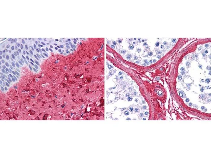
GeneTex anti collagen III antibody (GTX26580, 1:400, 45 min RT) showed strong staining in FFPE sections of human skin(left, dermis) with moderate to strong red staining and testis (right) where strong staining was observed within connective tissue between seminiferous tubules. The antibody showed strong extracellular staining within connective tissues across many organs with minimal background staining. Slides were steamed in 0.01 M sodium citrate buffer, pH 6.0 at 99-100oC - 20 minutes for antigen retrieval.
GeneTex anti collagen III antibody (GTX26580, 1:400, 45 min RT) showed strong staining in FFPE sections of human skin(left, dermis) with moderate to strong red staining and testis (right) where strong staining was observed within connective tissue between seminiferous tubules. The antibody showed strong extracellular staining within connective tissues across many organs with minimal background staining. Slides were steamed in 0.01 M sodium citrate buffer, pH 6.0 at 99-100oC - 20 minutes for antigen retrieval.
Collagen III antibody (Biotin)
GTX26580
ApplicationsImmunoPrecipitation, Western Blot, ELISA, ImmunoHistoChemistry, ImmunoHistoChemistry Paraffin
Product group Antibodies
ReactivityBovine, Human
TargetCOL3A1
Overview
- SupplierGeneTex
- Product NameCollagen III antibody (Biotin)
- Delivery Days Customer9
- Application Supplier NoteWB: 1:1000-1:5000. IHC-P: 1:200-1:1000. IP: 1:100. ELISA: 1:10000-1:50000. *Optimal dilutions/concentrations should be determined by the researcher.Not tested in other applications.
- ApplicationsImmunoPrecipitation, Western Blot, ELISA, ImmunoHistoChemistry, ImmunoHistoChemistry Paraffin
- CertificationResearch Use Only
- ClonalityPolyclonal
- Concentration1 mg/ml
- ConjugateBiotin
- Gene ID1281
- Target nameCOL3A1
- Target descriptioncollagen type III alpha 1 chain
- Target synonymsEDS4A, EDSVASC, PMGEDSV, collagen alpha-1(III) chain, Ehlers-Danlos syndrome type IV, autosomal dominant, alpha-1 type III collagen, alpha1 (III) collagen, collagen, fetal, collagen, type III, alpha 1
- HostRabbit
- IsotypeIgG
- Protein IDP02461
- Protein NameCollagen alpha-1(III) chain
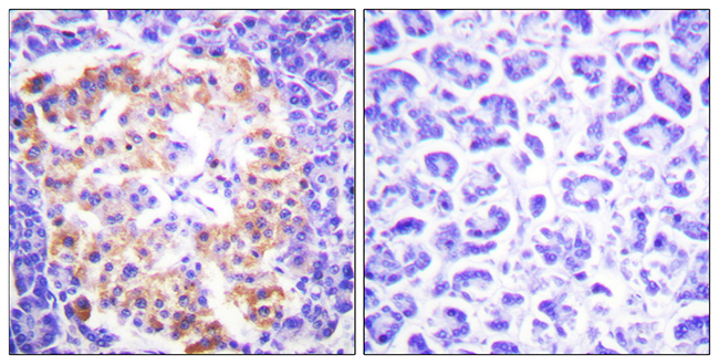
- Scientific DescriptionThis antibody is well suited to detect extracellular matrix proteins in normal as well as disease state tissues. Disruption of tissue organization is the hallmark of neoplasia. Malignant lesions can be distinguished from benign by examining the breakdown of basement membranes and loss of 3-dimensional architecture. Malignant cells are presumed to use matrix metalloproteases to degrade barriers created by the extracellular matrix, which then allows metastasis to occur. Collagenases, stomelysins and gelatinases can collectively degrade all of the various components of the extracellular matrix, including fibrillar and non-fibrillar collagens and basement membrane glycoproteins.
- ReactivityBovine, Human
- Storage Instruction-20°C or -80°C,2°C to 8°C
- UNSPSC41116161
Datasheet
Related products
Product group Antibodies
ApplicationsImmunoFluorescence, ELISA, ImmunoHistoChemistry
ReactivityHuman, Mouse, Rat
- SizePrice

Product group Antibodies
Anti-Collagen III/COL3A1 Antibody Picoband(r)A00788-3-CARRIER-FREE
ApplicationsFlow Cytometry, Western Blot, ELISA, ImmunoHistoChemistry
ReactivityHuman, Mouse, Rat
TargetCOL3A1
- SizePrice
Product group Antibodies
Anti-COL3A1 Antibody144-03795
ApplicationsWestern Blot
ReactivityHuman, Mouse
TargetCOL3A1
- SizePrice
Product group Antibodies

ApplicationsImmunoFluorescence, Western Blot, ImmunoHistoChemistry, ImmunoHistoChemistry Paraffin
ReactivityHuman, Mouse, Rat
TargetCOL3A1
- SizePrice

Product group Antibodies
References
Collagen III Polyclonal AntibodyBS-0549R
ApplicationsImmunoFluorescence, Western Blot, ImmunoCytoChemistry, ImmunoHistoChemistry, ImmunoHistoChemistry Frozen, ImmunoHistoChemistry Paraffin
ReactivityHuman
TargetCOL3A1
- SizePrice
Product group Antibodies
COL3A1 Monoclonal AntibodyCSB-MA000282
ApplicationsWestern Blot, ELISA
ReactivityHuman, Mouse, Rat
TargetCOL3A1
- SizePrice
Product group Antibodies

Procollagen Type III, human, bovineCO23311-0.1
ApplicationsImmunoFluorescence, Western Blot, ELISA, ImmunoHistoChemistry, ImmunoHistoChemistry Paraffin, RadioImmunoAssay
ReactivityBovine, Human, Porcine
TargetCOL3A1
- SizePrice
Product group Antibodies
ApplicationsFlow Cytometry
TargetCOL3A1
- SizePrice
![IHC-Fr analysis of rat skin tissue using GTX26310 Collagen III antibody [FH-7A] at 1:8,000.](https://www.genetex.com/upload/website/prouct_img/normal/GTX26310/GTX26310_20170605_IHC-Fr_w_23060722_325.webp)
Product group Antibodies
References
Collagen III antibody [FH-7A]GTX26310
ApplicationsDot Blot, Western Blot, ELISA, ImmunoHistoChemistry, ImmunoHistoChemistry Frozen, ImmunoHistoChemistry Paraffin
ReactivityHuman, Rabbit, Rat
TargetCOL3A1
- SizePrice