

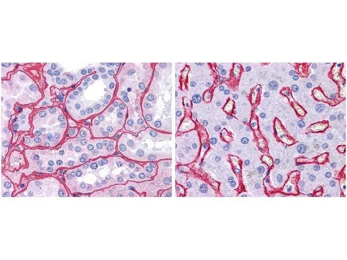
GeneTex anti collagen IV antibody (GTX26586, 1:400, 45 min RT) showed strong staining in FFPE sections of human kidney (Left) with strong red staining observed in glomeruli and liver (Right) with strong staining in sinusoids. Staining for both tissues was consistent with a basement membrane distribution. Slides were steamed in 0.01 M sodium citrate buffer, pH 6.0 at 99-100oC - 20 minutes for antigen retrieval.
GeneTex anti collagen IV antibody (GTX26586, 1:400, 45 min RT) showed strong staining in FFPE sections of human kidney (Left) with strong red staining observed in glomeruli and liver (Right) with strong staining in sinusoids. Staining for both tissues was consistent with a basement membrane distribution. Slides were steamed in 0.01 M sodium citrate buffer, pH 6.0 at 99-100oC - 20 minutes for antigen retrieval.
Collagen IV antibody
GTX26586
ApplicationsDot Blot, ImmunoFluorescence, ImmunoPrecipitation, Western Blot, ImmunoCytoChemistry, ImmunoHistoChemistry, ImmunoHistoChemistry Paraffin, Other Application
Product group Antibodies
ReactivityBovine, Human
TargetCOL4A1
Overview
- SupplierGeneTex
- Product NameCollagen IV antibody
- Delivery Days Customer9
- Application Supplier NoteWB: 1:1000-1:10000. IHC-P: 1:50-1:200. IP: 1:100. *Optimal dilutions/concentrations should be determined by the researcher.Not tested in other applications.
- ApplicationsDot Blot, ImmunoFluorescence, ImmunoPrecipitation, Western Blot, ImmunoCytoChemistry, ImmunoHistoChemistry, ImmunoHistoChemistry Paraffin, Other Application
- CertificationResearch Use Only
- ClonalityPolyclonal
- Concentration1.17 mg/ml
- ConjugateUnconjugated
- Gene ID1282
- Target nameCOL4A1
- Target descriptioncollagen type IV alpha 1 chain
- Target synonymsBSVD, BSVD1, COL4A1s, PADMAL, RATOR, collagen alpha-1(IV) chain, COL4A1 NC1 domain, arresten, collagen IV, alpha-1 polypeptide, collagen of basement membrane, alpha-1 chain
- HostRabbit
- IsotypeIgG
- Protein IDP02462
- Protein NameCollagen alpha-1(IV) chain
- Scientific DescriptionThis antibody is well suited to detect extracellular matrix proteins in normal as well as disease state tissues. Disruption of tissue organization is the hallmark of neoplasia. Malignant lesions can be distinguished from benign by examining the breakdown of basement membranes and loss of 3-dimensional architecture. Malignant cells are presumed to use matrix metalloproteases to degrade barriers created by the extracellular matrix, which then allows metastasis to occur. Collagenases, stomelysins and gelatinases can collectively degrade all of the various components of the extracellular matrix, including fibrillar and non-fibrillar collagens and basement membrane glycoproteins.
- ReactivityBovine, Human
- Storage Instruction-20°C or -80°C,2°C to 8°C
- UNSPSC41116161
References
- A ROCK inhibitor suppresses the transforming growth factor-beta-2-induced endothelial-mesenchymal transition in Schlemms canal endothelial cells.Read this paper
- Injection of hybrid 3D spheroids composed of podocytes, mesenchymal stem cells, and vascular endothelial cells into the renal cortex improves kidney function and replenishes glomerular podocytes. Yang WY et al., 2021 May, Bioeng Transl MedRead this paper
- Investigating the effect of chitosan/ polycaprolactone blends in differentiation of corneal endothelial cells and extracellular matrix compositions. Wang YH et al., 2019 Aug, Exp Eye ResRead this paper
Datasheet
Related products
Product group Antibodies
ApplicationsWestern Blot, ELISA, ImmunoHistoChemistry
ReactivityHuman, Mouse
- SizePrice
Product group Antibodies
Anti-Collagen Type IV Antibody118-10009
ApplicationsWestern Blot, ELISA, ImmunoHistoChemistry
ReactivityHuman
- SizePrice
Product group Antibodies

ApplicationsImmunoFluorescence, ImmunoHistoChemistry, ImmunoHistoChemistry Paraffin
ReactivityHuman, Mouse, Rat
TargetCOL4A1
- SizePrice

Product group Antibodies
COL4A1 Monoclonal AntibodyCSB-MA145731
ApplicationsELISA, ImmunoHistoChemistry
ReactivityHuman, Mouse, Rat
TargetCOL4A1
- SizePrice
Product group Antibodies

ApplicationsWestern Blot, ImmunoHistoChemistry, ImmunoHistoChemistry Frozen, ImmunoHistoChemistry Paraffin
ReactivityGoat, Human
TargetCOL4A1
- SizePrice
Product group Antibodies
Col4A1 Polyclonal AntibodyCAC07119
ApplicationsImmunoFluorescence, ELISA, ImmunoHistoChemistry
TargetCOL4A1
- SizePrice
Product group Antibodies
References
Collagen IV Polyclonal AntibodyBS-4595R
ApplicationsFlow Cytometry, ImmunoFluorescence, Western Blot, ELISA, ImmunoCytoChemistry, ImmunoHistoChemistry, ImmunoHistoChemistry Frozen, ImmunoHistoChemistry Paraffin
ReactivityHuman
TargetCOL4A1
- SizePrice
Product group Antibodies
References
Collagen IV antibodyGTX19808
ApplicationsImmunoFluorescence, ELISA, ImmunoCytoChemistry, ImmunoHistoChemistry, ImmunoHistoChemistry Paraffin, RadioImmunoAssay
ReactivityMouse
TargetCOL4A1
- SizePrice
Product group Antibodies
Collagen IV antibody [ZR108]GTX01655
ApplicationsImmunoHistoChemistry, ImmunoHistoChemistry Paraffin
ReactivityHuman
TargetCOL4A1
- SizePrice