

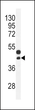
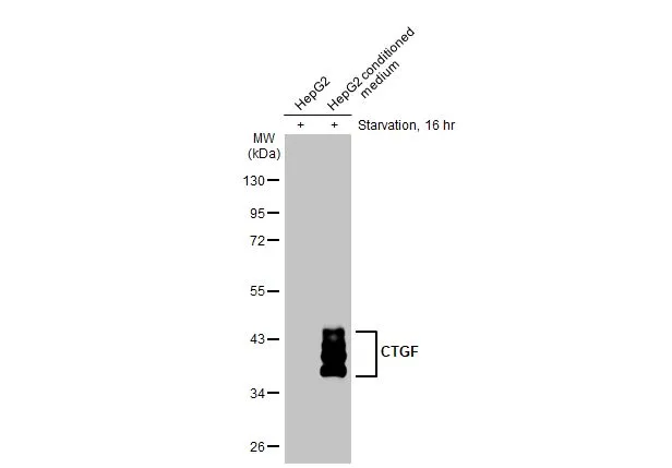
WB analysis of mouse bladder tissue lysate (35ug/lane) using GTX81297 CTGF antibody, Internal.

WB analysis of mouse bladder tissue lysate (35ug/lane) using GTX81297 CTGF antibody, Internal.
CTGF antibody, Internal
GTX81297
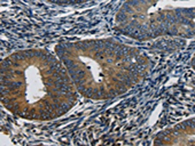
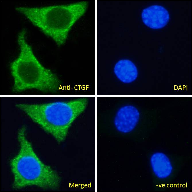
ApplicationsWestern Blot, ImmunoHistoChemistry, ImmunoHistoChemistry Paraffin
Product group Antibodies
ReactivityHuman, Mouse
TargetCCN2
Overview
- SupplierGeneTex
- Product NameCTGF antibody, Internal
- Delivery Days Customer9
- Application Supplier NoteWB: 1:1000. IHC-P: 1:10-1:50. *Optimal dilutions/concentrations should be determined by the researcher.Not tested in other applications.
- ApplicationsWestern Blot, ImmunoHistoChemistry, ImmunoHistoChemistry Paraffin
- CertificationResearch Use Only
- ClonalityPolyclonal
- ConjugateUnconjugated
- Gene ID1490
- Target nameCCN2
- Target descriptioncellular communication network factor 2
- Target synonymsCTGF, HCS24, IBP-8, IGFBP8, KMD, NOV2, SEMDLSL, CCN family member 2, IGF-binding protein 8, connective tissue growth factor, hypertrophic chondrocyte-specific protein 24, insulin-like growth factor-binding protein 8
- HostRabbit
- IsotypeIgG
- Protein IDP29279
- Protein NameCCN family member 2
- Scientific DescriptionThe protein encoded by this gene is a mitogen that is secreted by vascular endothelial cells. The encoded protein plays a role in chondrocyte proliferation and differentiation, cell adhesion in many cell types, and is related to platelet-derived growth factor. Certain polymorphisms in this gene have been linked with a higher incidence of systemic sclerosis. [provided by RefSeq, Nov 2009]
- ReactivityHuman, Mouse
- Storage Instruction-20°C or -80°C,2°C to 8°C
- UNSPSC41116161
Datasheet
Related products
Product group Antibodies
CTGF AntibodyCSB-PA060417
ApplicationsWestern Blot, ELISA, ImmunoHistoChemistry
ReactivityHuman, Mouse, Rat
TargetCCN2
- SizePrice

Product group Antibodies
Anti-CTGF AntibodyAMAB91366
ApplicationsWestern Blot, ImmunoHistoChemistry
ReactivityHuman, Mouse, Rat
TargetCCN2
- SizePrice
Product group Antibodies

Anti-CTGF [HL2180 [17-73-D6]]AB02551-10.0-BT
ApplicationsWestern Blot, ELISA, ImmunoHistoChemistry
ReactivityHuman, Rabbit
TargetCCN2
- SizePrice
Product group Antibodies
Anti-CTGF AntibodyA285947
ApplicationsImmunoFluorescence, ELISA, ImmunoHistoChemistry
ReactivityHuman, Mouse, Rat
- SizePrice
Product group Antibodies

ApplicationsImmunoFluorescence, ELISA, ImmunoHistoChemistry
ReactivityCanine, Human, Mouse, Rat
TargetCCN2
- SizePrice
Product group Antibodies

CTGF AntibodyLS-C402210
ApplicationsWestern Blot, ELISA, ImmunoHistoChemistry
ReactivityHuman
TargetCCN2
- SizePrice
Product group Antibodies
ApplicationsImmunoPrecipitation, Western Blot, ImmunoCytoChemistry, ImmunoHistoChemistry
ReactivityMouse, Rat
TargetCCN2
- SizePrice

Product group Antibodies
Anti-CTGF Antibody Picoband(r)PB9541-CARRIER-FREE
ApplicationsWestern Blot, ImmunoHistoChemistry
ReactivityHuman, Rat
TargetCCN2
- SizePrice
Product group Antibodies
CTGF antibodyGTX04889
ApplicationsImmunoFluorescence, Western Blot, ImmunoCytoChemistry
ReactivityHuman
TargetCCN2
- SizePrice
Product group Antibodies
CTGF antibodyGTX124232
ApplicationsWestern Blot, ImmunoHistoChemistry, ImmunoHistoChemistry Frozen, ImmunoHistoChemistry Paraffin
ReactivityBovine, Human, Mouse, Primate, Rat
TargetCCN2
- SizePrice