

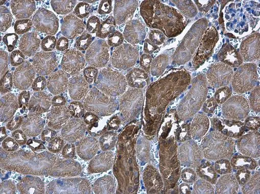
Cytochrome C antibody [GT577] detects Cytochrome C protein at cytoplasm in mouse kidney by immunohistochemical analysis. Sample: Paraffin-embedded mouse kidney. Cytochrome C antibody [GT577] (GTX633691) diluted at 1:200.
Antigen Retrieval: Citrate buffer, pH 6.0, 15 min
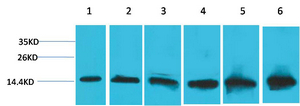
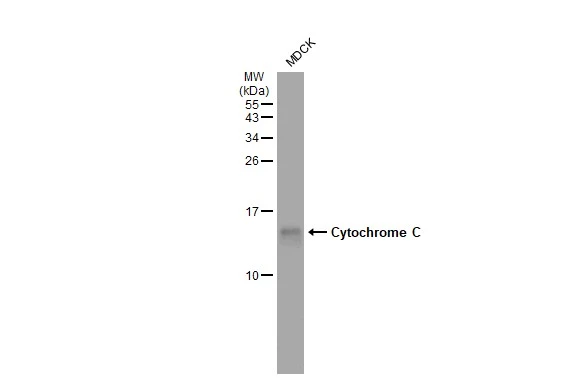
 was separated by 15% SDS-PAGE, and the membrane was blotted with Cytochrome C antibody (GTX633691) diluted at 1:3000.")
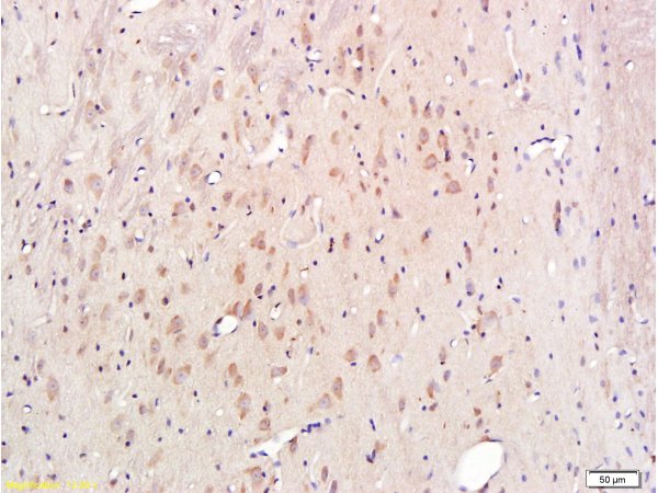
![Cytochrome C antibody [GT577] detects Cytochrome C protein at cytoplasm in rat muscle by immunohistochemical analysis. Sample: Paraffin-embedded rat muscle. Cytochrome C antibody [GT577] (GTX633691) diluted at 1:200.
Antigen Retrieval: Citrate buffer, pH 6.0, 15 min](https://www.genetex.com/upload/website/prouct_img/normal/GTX633691/GTX633691_42632_20161219_IHC-P_R_w_23061202_971.webp "Cytochrome C antibody [GT577] detects Cytochrome C protein at cytoplasm in rat muscle by immunohistochemical analysis. Sample: Paraffin-embedded rat muscle. Cytochrome C antibody [GT577] (GTX633691) diluted at 1:200.
Antigen Retrieval: Citrate buffer, pH 6.0, 15 min")
![Whole cell extract (30 μg) was separated by 15% SDS-PAGE, and the membrane was blotted with Cytochrome C antibody [GT577] (GTX633691) diluted at 1:500.](https://www.genetex.com/upload/website/prouct_img/normal/GTX633691/GTX633691_42632_20161027_WB_w_23061202_482.webp "Whole cell extract (30 μg) was separated by 15% SDS-PAGE, and the membrane was blotted with Cytochrome C antibody [GT577] (GTX633691) diluted at 1:500.")
Cytochrome C antibody [GT577] detects Cytochrome C protein at cytoplasm in mouse kidney by immunohistochemical analysis. Sample: Paraffin-embedded mouse kidney. Cytochrome C antibody [GT577] (GTX633691) diluted at 1:200.
Antigen Retrieval: Citrate buffer, pH 6.0, 15 min
Cytochrome C antibody [GT577]
GTX633691
ApplicationsWestern Blot, ImmunoHistoChemistry, ImmunoHistoChemistry Paraffin
Product group Antibodies
ReactivityHuman, Mouse, Rat
TargetCYCS
Overview
- SupplierGeneTex
- Product NameCytochrome C antibody [GT577]
- Delivery Days Customer9
- Application Supplier NoteWB: 1:500-1:3000. IHC-P: 1:100-1:1000. *Optimal dilutions/concentrations should be determined by the researcher.Not tested in other applications.
- ApplicationsWestern Blot, ImmunoHistoChemistry, ImmunoHistoChemistry Paraffin
- CertificationResearch Use Only
- ClonalityMonoclonal
- Clone IDGT577
- Concentration1 mg/ml
- ConjugateUnconjugated
- Gene ID54205
- Target nameCYCS
- Target descriptioncytochrome c, somatic
- Target synonymsCYC, HCS, THC4, cytochrome c
- HostMouse
- IsotypeIgA
- Protein IDP99999
- Protein NameCytochrome c
- Scientific DescriptionThis gene encodes cytochrome c, a component of the electron transport chain in mitochondria. The heme group of cytochrome c accepts electrons from the b-c1 complex and transfers electrons to the cytochrome oxidase complex. Cytochrome c is also involved in initiation of apoptosis. Upon release of cytochrome c to the cytoplasm, the protein binds apoptotic protease activating factor which activates the apoptotic initiator procaspase 9. Many cytochrome c pseudogenes exist, scattered throughout the human genome. [provided by RefSeq]
- ReactivityHuman, Mouse, Rat
- Storage Instruction-20°C or -80°C,2°C to 8°C
- UNSPSC41116161
Datasheet
Related products
Product group Antibodies
Anti-CYCS AntibodyA36381
ApplicationsWestern Blot, ImmunoHistoChemistry
ReactivityHuman, Mouse, Rat
- SizePrice
Product group Antibodies
Anti-CYCS Antibody144-60334
ApplicationsImmunoFluorescence, Western Blot, ImmunoHistoChemistry
ReactivityHuman, Mouse, Rat
TargetCYCS
- SizePrice
Product group Antibodies
References
Cytochrome C Polyclonal AntibodyBS-0013R
ApplicationsFlow Cytometry, ImmunoFluorescence, Western Blot, ELISA, ImmunoCytoChemistry, ImmunoHistoChemistry, ImmunoHistoChemistry Frozen, ImmunoHistoChemistry Paraffin
ReactivityBovine, Equine, Mouse, Porcine, Rat
TargetCYCS
- SizePrice
Product group Antibodies
CYCS Monoclonal AntibodyCSB-MA080196
ApplicationsWestern Blot, ELISA, ImmunoHistoChemistry
ReactivityHuman, Mouse, Rat
TargetCYCS
- SizePrice
Product group Antibodies
Cycs Polyclonal AntibodyCAC07497
ApplicationsWestern Blot, ELISA, ImmunoHistoChemistry
ReactivityMouse, Rat
TargetCYCS
- SizePrice
![FACS analysis of PFA fixed HeLa cells using GTX13575 Cytochrome C antibody [7H8.2C12]. Blue : Primary antibody Red : Isotype control](https://www.genetex.com/upload/website/prouct_img/normal/GTX13575/GTX13575_20200115_FACS_1678_1021_w_23060620_766.webp)
Product group Antibodies
References
Cytochrome C antibody [7H8.2C12]GTX13575
ApplicationsFlow Cytometry, Western Blot, ImmunoHistoChemistry, ImmunoHistoChemistry Paraffin
ReactivityAmphibian, Avian, Canine, Drosophila, Equine, Human, Mouse, Porcine, Rat
TargetCYCS
- SizePrice
Product group Antibodies
ApplicationsWestern Blot
ReactivityGoat, Human, Monkey, Mouse, Rat, Sheep
TargetCYCS
- SizePrice
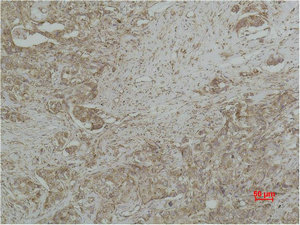
![IHC-P analysis of human liver tissue section using GTX02626 Cytochrome C antibody [CYCS/3128R].](https://www.genetex.com/upload/website/prouct_img/normal/GTX02626/GTX02626_20210319_IHC-P_w_23053122_255.webp)
Product group Antibodies
ApplicationsFlow Cytometry, ImmunoFluorescence, Western Blot, ImmunoCytoChemistry, ImmunoHistoChemistry, ImmunoHistoChemistry Paraffin
ReactivityAmphibian, Avian, Canine, Drosophila, Equine, Human, Mouse, Rat
TargetCYCS
- SizePrice
Product group Antibodies
Cytochrome C antibodyGTX108585
ApplicationsImmunoFluorescence, Western Blot, ImmunoCytoChemistry, ImmunoHistoChemistry, ImmunoHistoChemistry Frozen, ImmunoHistoChemistry Paraffin
ReactivityCanine, Drosophila, Human, Mouse, Rat
TargetCYCS
- SizePrice
![Mouse tissue extract (50 μg) was separated by 15% SDS-PAGE, and the membrane was blotted with Cytochrome C antibody [HL2205] (GTX638209) diluted at 1:2000. The HRP-conjugated anti-rabbit IgG antibody (GTX213110-01) was used to detect the primary antibody.](https://www.genetex.com/upload/website/prouct_img/normal/GTX638209/GTX638209_T-44942_20230210_WB_M_brain_23021401_315.webp)
Product group Antibodies
Cytochrome C antibody [HL2205]GTX638209
ApplicationsImmunoFluorescence, Western Blot, ImmunoCytoChemistry, ImmunoHistoChemistry, ImmunoHistoChemistry Paraffin
ReactivityFeline, Human, Mouse, Rat
TargetCYCS
- SizePrice