

Mouse tissue extract (50 μg) was separated by 15% SDS-PAGE, and the membrane was blotted with Cytochrome C antibody [HL2205] (GTX638209) diluted at 1:2000. The HRP-conjugated anti-rabbit IgG antibody (GTX213110-01) was used to detect the primary antibody.
![Cytochrome C antibody [HL2205] detects Cytochrome C protein at mitochondria by immunohistochemical analysis. Sample: Paraffin-embedded human lung cancer. Cytochrome C stained by Cytochrome C antibody [HL2205] (GTX638209) diluted at 1:100. Antigen Retrieval: Citrate buffer, pH 6.0, 15 min](https://www.genetex.com/upload/website/prouct_img/normal/GTX638209/GTX638209_T-44942_20230303_IHC-P_23031402_575.webp "Cytochrome C antibody [HL2205] detects Cytochrome C protein at mitochondria by immunohistochemical analysis. Sample: Paraffin-embedded human lung cancer. Cytochrome C stained by Cytochrome C antibody [HL2205] (GTX638209) diluted at 1:100. Antigen Retrieval: Citrate buffer, pH 6.0, 15 min")
![Cytochrome C antibody [HL2205] detects Cytochrome C protein at mitochondria by immunofluorescent analysis. Sample: HeLa cells were fixed in ice-cold MeOH for 5 min. Green: Cytochrome C stained by Cytochrome C antibody [HL2205] (GTX638209) diluted at 1:500. Red: alpha Tubulin, a cytoskeleton marker, stained by alpha Tubulin antibody [GT114] (GTX628802) diluted at 1:1000. Blue: Fluoroshield with DAPI (GTX30920).](https://www.genetex.com/upload/website/prouct_img/normal/GTX638209/GTX638209_T-44942_20230331_ICC_IF_23041023_864.webp "Cytochrome C antibody [HL2205] detects Cytochrome C protein at mitochondria by immunofluorescent analysis. Sample: HeLa cells were fixed in ice-cold MeOH for 5 min. Green: Cytochrome C stained by Cytochrome C antibody [HL2205] (GTX638209) diluted at 1:500. Red: alpha Tubulin, a cytoskeleton marker, stained by alpha Tubulin antibody [GT114] (GTX628802) diluted at 1:1000. Blue: Fluoroshield with DAPI (GTX30920).")
![Various whole cell extracts (30 μg) were separated by 15% SDS-PAGE, and the membrane was blotted with Cytochrome C antibody [HL2205] (GTX638209) diluted at 1:1000. The HRP-conjugated anti-rabbit IgG antibody (GTX213110-01) was used to detect the primary antibody. Corresponding RNA expression data for the same cell lines are based on Human Protein Atlas program.](https://www.genetex.com/upload/website/prouct_img/normal/GTX638209/GTX638209_45012_20230421_WB_TPM_watermark_23042500_470.webp "Various whole cell extracts (30 μg) were separated by 15% SDS-PAGE, and the membrane was blotted with Cytochrome C antibody [HL2205] (GTX638209) diluted at 1:1000. The HRP-conjugated anti-rabbit IgG antibody (GTX213110-01) was used to detect the primary antibody. Corresponding RNA expression data for the same cell lines are based on Human Protein Atlas program.")
![Whole cell extract (50 μg) was separated by 15% SDS-PAGE, and the membrane was blotted with Cytochrome C antibody [HL2205] (GTX638209) diluted at 1:1000. The HRP-conjugated anti-rabbit IgG antibody (GTX213110-01) was used to detect the primary antibody, and the signal was developed with Trident ECL plus-Enhanced.](https://www.genetex.com/upload/website/prouct_img/normal/GTX638209/GTX638209_45012_20230519_WB_R_23053001_233.webp "Whole cell extract (50 μg) was separated by 15% SDS-PAGE, and the membrane was blotted with Cytochrome C antibody [HL2205] (GTX638209) diluted at 1:1000. The HRP-conjugated anti-rabbit IgG antibody (GTX213110-01) was used to detect the primary antibody, and the signal was developed with Trident ECL plus-Enhanced.")
![Rat tissue extract (50 μg) was separated by 15% SDS-PAGE, and the membrane was blotted with Cytochrome C antibody [HL2205] (GTX638209) diluted at 1:1000. The HRP-conjugated anti-rabbit IgG antibody (GTX213110-01) was used to detect the primary antibody.](https://www.genetex.com/upload/website/prouct_img/normal/GTX638209/GTX638209_45012_20230519_WB_R_brain_23053001_262.webp "Rat tissue extract (50 μg) was separated by 15% SDS-PAGE, and the membrane was blotted with Cytochrome C antibody [HL2205] (GTX638209) diluted at 1:1000. The HRP-conjugated anti-rabbit IgG antibody (GTX213110-01) was used to detect the primary antibody.")
![Non-transfected (–) and transfected (+) 293T whole cell extracts (30 μg) were separated by 15% SDS-PAGE, and the membrane was blotted with Cytochrome C antibody [HL2205] (GTX638209) diluted at 1:1000. The HRP-conjugated anti-rabbit IgG antibody (GTX213110-01) was used to detect the primary antibody.](https://www.genetex.com/upload/website/prouct_img/normal/GTX638209/GTX638209_45012_20230707_WB_shRNA_watermark_23071300_650.webp "Non-transfected (–) and transfected (+) 293T whole cell extracts (30 μg) were separated by 15% SDS-PAGE, and the membrane was blotted with Cytochrome C antibody [HL2205] (GTX638209) diluted at 1:1000. The HRP-conjugated anti-rabbit IgG antibody (GTX213110-01) was used to detect the primary antibody.")
![Cytochrome C antibody [HL2205] detects Cytochrome C protein at mitochondria by immunohistochemical analysis. Sample: Paraffin-embedded cat kidney. Cytochrome C stained by Cytochrome C antibody [HL2205] (GTX638209) diluted at 1:100. Antigen Retrieval: Citrate buffer, pH 6.0, 15 min](https://www.genetex.com/upload/website/prouct_img/normal/GTX638209/GTX638209_45012_20230721_IHC-P_Cat_23073119_569.webp "Cytochrome C antibody [HL2205] detects Cytochrome C protein at mitochondria by immunohistochemical analysis. Sample: Paraffin-embedded cat kidney. Cytochrome C stained by Cytochrome C antibody [HL2205] (GTX638209) diluted at 1:100. Antigen Retrieval: Citrate buffer, pH 6.0, 15 min")
![Various whole cell extracts (30 μg) were separated by 15% SDS-PAGE, and the membrane was blotted with Cytochrome C antibody [HL2205] (GTX638209) diluted at 1:1000. The HRP-conjugated anti-rabbit IgG antibody (GTX213110-01) was used to detect the primary antibody.](https://www.genetex.com/upload/website/prouct_img/normal/GTX638209/GTX638209_45012_20241011_WB_24101600_763.webp "Various whole cell extracts (30 μg) were separated by 15% SDS-PAGE, and the membrane was blotted with Cytochrome C antibody [HL2205] (GTX638209) diluted at 1:1000. The HRP-conjugated anti-rabbit IgG antibody (GTX213110-01) was used to detect the primary antibody.")
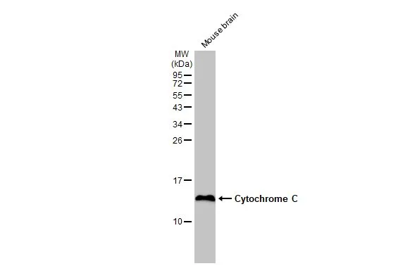
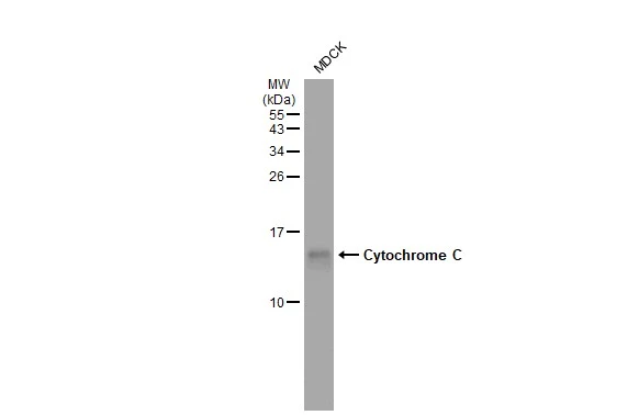
Mouse tissue extract (50 μg) was separated by 15% SDS-PAGE, and the membrane was blotted with Cytochrome C antibody [HL2205] (GTX638209) diluted at 1:2000. The HRP-conjugated anti-rabbit IgG antibody (GTX213110-01) was used to detect the primary antibody.
Cytochrome C antibody [HL2205]
GTX638209
ApplicationsImmunoFluorescence, Western Blot, ImmunoCytoChemistry, ImmunoHistoChemistry, ImmunoHistoChemistry Paraffin
Product group Antibodies
ReactivityFeline, Human, Mouse, Rat
TargetCYCS
Overview
- SupplierGeneTex
- Product NameCytochrome C antibody [HL2205]
- Delivery Days Customer9
- ApplicationsImmunoFluorescence, Western Blot, ImmunoCytoChemistry, ImmunoHistoChemistry, ImmunoHistoChemistry Paraffin
- CertificationResearch Use Only
- ClonalityMonoclonal
- Clone IDHL2205
- Concentration2 mg/ml
- ConjugateUnconjugated
- Gene ID54205
- Target nameCYCS
- Target descriptioncytochrome c, somatic
- Target synonymsCYC, HCS, THC4, cytochrome c
- HostRabbit
- IsotypeIgG
- Protein IDP99999
- Protein NameCytochrome c
- Scientific DescriptionThis gene encodes a small heme protein that functions as a central component of the electron transport chain in mitochondria. The encoded protein associates with the inner membrane of the mitochondrion where it accepts electrons from cytochrome b and transfers them to the cytochrome oxidase complex. This protein is also involved in initiation of apoptosis. Mutations in this gene are associated with autosomal dominant nonsyndromic thrombocytopenia. Numerous processed pseudogenes of this gene are found throughout the human genome.[provided by RefSeq, Jul 2010]
- ReactivityFeline, Human, Mouse, Rat
- Storage Instruction-20°C or -80°C,2°C to 8°C
- UNSPSC41116161
Datasheet
Related products
Product group Antibodies
Anti-CYCS AntibodyA36381
ApplicationsWestern Blot, ImmunoHistoChemistry
ReactivityHuman, Mouse, Rat
- SizePrice
Product group Antibodies
Anti-CYCS Antibody144-60334
ApplicationsImmunoFluorescence, Western Blot, ImmunoHistoChemistry
ReactivityHuman, Mouse, Rat
TargetCYCS
- SizePrice
Product group Antibodies
References
Cytochrome C Polyclonal AntibodyBS-0013R
ApplicationsFlow Cytometry, ImmunoFluorescence, Western Blot, ELISA, ImmunoCytoChemistry, ImmunoHistoChemistry, ImmunoHistoChemistry Frozen, ImmunoHistoChemistry Paraffin
ReactivityBovine, Equine, Mouse, Porcine, Rat
TargetCYCS
- SizePrice
Product group Antibodies
CYCS Monoclonal AntibodyCSB-MA080196
ApplicationsWestern Blot, ELISA, ImmunoHistoChemistry
ReactivityHuman, Mouse, Rat
TargetCYCS
- SizePrice
Product group Antibodies
Cycs Polyclonal AntibodyCAC07497
ApplicationsWestern Blot, ELISA, ImmunoHistoChemistry
ReactivityMouse, Rat
TargetCYCS
- SizePrice
![FACS analysis of PFA fixed HeLa cells using GTX13575 Cytochrome C antibody [7H8.2C12]. Blue : Primary antibody Red : Isotype control](https://www.genetex.com/upload/website/prouct_img/normal/GTX13575/GTX13575_20200115_FACS_1678_1021_w_23060620_766.webp)
Product group Antibodies
References
Cytochrome C antibody [7H8.2C12]GTX13575
ApplicationsFlow Cytometry, Western Blot, ImmunoHistoChemistry, ImmunoHistoChemistry Paraffin
ReactivityAmphibian, Avian, Canine, Drosophila, Equine, Human, Mouse, Porcine, Rat
TargetCYCS
- SizePrice
Product group Antibodies
ApplicationsWestern Blot
ReactivityGoat, Human, Monkey, Mouse, Rat, Sheep
TargetCYCS
- SizePrice
![IHC-P analysis of human liver tissue section using GTX02626 Cytochrome C antibody [CYCS/3128R].](https://www.genetex.com/upload/website/prouct_img/normal/GTX02626/GTX02626_20210319_IHC-P_w_23053122_255.webp)
Product group Antibodies
ApplicationsFlow Cytometry, ImmunoFluorescence, Western Blot, ImmunoCytoChemistry, ImmunoHistoChemistry, ImmunoHistoChemistry Paraffin
ReactivityAmphibian, Avian, Canine, Drosophila, Equine, Human, Mouse, Rat
TargetCYCS
- SizePrice
Product group Antibodies
Cytochrome C antibodyGTX108585
ApplicationsImmunoFluorescence, Western Blot, ImmunoCytoChemistry, ImmunoHistoChemistry, ImmunoHistoChemistry Frozen, ImmunoHistoChemistry Paraffin
ReactivityCanine, Drosophila, Human, Mouse, Rat
TargetCYCS
- SizePrice
![Cytochrome C antibody [GT577] detects Cytochrome C protein at cytoplasm in mouse kidney by immunohistochemical analysis. Sample: Paraffin-embedded mouse kidney. Cytochrome C antibody [GT577] (GTX633691) diluted at 1:200.
Antigen Retrieval: Citrate buffer, pH 6.0, 15 min](https://www.genetex.com/upload/website/prouct_img/normal/GTX633691/GTX633691_42632_20161219_IHC-P_M_w_23061202_371.webp)
Product group Antibodies
Cytochrome C antibody [GT577]GTX633691
ApplicationsWestern Blot, ImmunoHistoChemistry, ImmunoHistoChemistry Paraffin
ReactivityHuman, Mouse, Rat
TargetCYCS
- SizePrice