

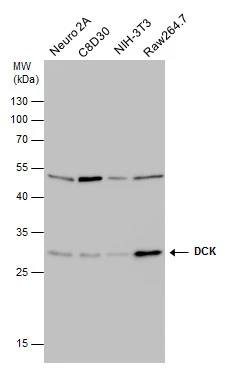
DCK antibody [GT10412] detects DCK protein by western blot analysis. Various whole cell extracts (30 μg) were separated by 12% SDS-PAGE, and the membrane was blotted with DCK antibody [GT10412] (GTX632065) diluted at a dilution of 1:500.
 were separated by 12% SDS-PAGE, and the membrane was blotted with DCK antibody (GTX632065) diluted at a dilution of 1:500.")
![DCK antibody [GT10412] detects DCK protein by western blot analysis. Various whole cell extracts (30 μg) were separated by 12% SDS-PAGE, and the membrane was blotted with DCK antibody [GT10412] (GTX632065) diluted at a dilution of 1:500.](https://www.genetex.com/upload/website/prouct_img/normal/GTX632065/GTX632065_41967_20150515_WB_R_w_23061202_551.webp "DCK antibody [GT10412] detects DCK protein by western blot analysis. Various whole cell extracts (30 μg) were separated by 12% SDS-PAGE, and the membrane was blotted with DCK antibody [GT10412] (GTX632065) diluted at a dilution of 1:500.")
![DCK antibody [GT10412] detects DCK protein at nucleus and cytoplasm in human lung cancer by immunohistochemical analysis. Sample: Paraffin-embedded human lung cancer. DCK antibody [GT10412] (GTX632065) diluted at 1:1000.
Antigen Retrieval: Citrate buffer, pH 6.0, 15 min](https://www.genetex.com/upload/website/prouct_img/normal/GTX632065/GTX632065_41967_20150409_IHC_2_w_23061202_811.webp "DCK antibody [GT10412] detects DCK protein at nucleus and cytoplasm in human lung cancer by immunohistochemical analysis. Sample: Paraffin-embedded human lung cancer. DCK antibody [GT10412] (GTX632065) diluted at 1:1000.
Antigen Retrieval: Citrate buffer, pH 6.0, 15 min")
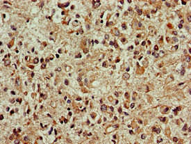
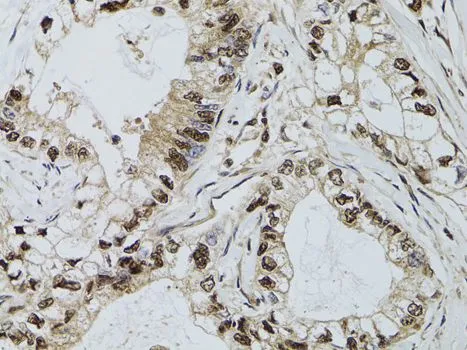
![DCK antibody [GT10412] detects DCK protein at nucleus and cytoplasm in human colon carcinoma by immunohistochemical analysis. Sample: Paraffin-embedded human colon carcinoma. DCK antibody [GT10412] (GTX632065) diluted at 1:1000.
Antigen Retrieval: Citrate buffer, pH 6.0, 15 min](https://www.genetex.com/upload/website/prouct_img/normal/GTX632065/GTX632065_41967_20150409_IHC_w_23061202_418.webp "DCK antibody [GT10412] detects DCK protein at nucleus and cytoplasm in human colon carcinoma by immunohistochemical analysis. Sample: Paraffin-embedded human colon carcinoma. DCK antibody [GT10412] (GTX632065) diluted at 1:1000.
Antigen Retrieval: Citrate buffer, pH 6.0, 15 min")
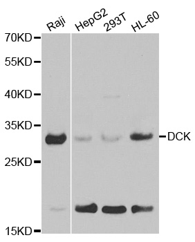
![Various whole cell extracts (30 μg) were separated by 12% SDS-PAGE, and the membrane was blotted with DCK antibody [GT10412] (GTX632065) diluted at 1:500.](https://www.genetex.com/upload/website/prouct_img/normal/GTX632065/GTX632065_41967_20160728_WB_w_23061202_779.webp "Various whole cell extracts (30 μg) were separated by 12% SDS-PAGE, and the membrane was blotted with DCK antibody [GT10412] (GTX632065) diluted at 1:500.")
![Immunoprecipitation of DCK protein from HCT-116 whole cell extract using 5 μg of DCK antibody [GT10412] (GTX632065) or DCK antibody (GTX102800). Western blot analysis was performed using DCK antibody [GT10412] (GTX632065) diluted at 1:500. EasyBlot HRP-conjugated anti mouse IgG antibody (GTX221667-01) was used to detect the primary antibody.](https://www.genetex.com/upload/website/prouct_img/normal/GTX632065/GTX632065_41967_20170428_IP_w_23061202_585.webp "Immunoprecipitation of DCK protein from HCT-116 whole cell extract using 5 μg of DCK antibody [GT10412] (GTX632065) or DCK antibody (GTX102800). Western blot analysis was performed using DCK antibody [GT10412] (GTX632065) diluted at 1:500. EasyBlot HRP-conjugated anti mouse IgG antibody (GTX221667-01) was used to detect the primary antibody.")
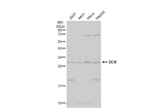
![Non-transfected (–) and transfected (+) 293T whole cell extracts (30 μg) were separated by 12% SDS-PAGE, and the membrane was blotted with DCK antibody [GT10412] (GTX632065) diluted at 1:5000. The HRP-conjugated anti-mouse IgG antibody (GTX213111-01) was used to detect the primary antibody.](https://www.genetex.com/upload/website/prouct_img/normal/GTX632065/GTX632065_41967_20180817_WB_B_w_23061202_944.webp "Non-transfected (–) and transfected (+) 293T whole cell extracts (30 μg) were separated by 12% SDS-PAGE, and the membrane was blotted with DCK antibody [GT10412] (GTX632065) diluted at 1:5000. The HRP-conjugated anti-mouse IgG antibody (GTX213111-01) was used to detect the primary antibody.")
![DCK antibody [GT10412] detects DCK protein at nucleus by immunofluorescent analysis. Sample: HCT 116 cells were fixed in 4% paraformaldehyde at RT for 15 min. Red: DCK protein stained by DCK antibody [GT10412] (GTX632065) diluted at 1:500. Blue: Hoechst 33342 staining.](https://www.genetex.com/upload/website/prouct_img/normal/GTX632065/GTX632065_41967_20160301_IFA_w_23061202_410.webp "DCK antibody [GT10412] detects DCK protein at nucleus by immunofluorescent analysis. Sample: HCT 116 cells were fixed in 4% paraformaldehyde at RT for 15 min. Red: DCK protein stained by DCK antibody [GT10412] (GTX632065) diluted at 1:500. Blue: Hoechst 33342 staining.")
DCK antibody [GT10412] detects DCK protein by western blot analysis. Various whole cell extracts (30 μg) were separated by 12% SDS-PAGE, and the membrane was blotted with DCK antibody [GT10412] (GTX632065) diluted at a dilution of 1:500.
DCK antibody [GT10412]
GTX632065
ApplicationsImmunoFluorescence, ImmunoPrecipitation, Western Blot, ImmunoCytoChemistry, ImmunoHistoChemistry, ImmunoHistoChemistry Paraffin
Product group Antibodies
ReactivityHuman, Mouse, Rat
TargetDCK
Overview
- SupplierGeneTex
- Product NameDCK antibody [GT10412]
- Delivery Days Customer9
- Application Supplier NoteWB: 1:1000-1:10000. ICC/IF: 1:100-1:1000. IHC-P: 1:100-1:1000. IP: 1:100-1:500. *Optimal dilutions/concentrations should be determined by the researcher.Not tested in other applications.
- ApplicationsImmunoFluorescence, ImmunoPrecipitation, Western Blot, ImmunoCytoChemistry, ImmunoHistoChemistry, ImmunoHistoChemistry Paraffin
- CertificationResearch Use Only
- ClonalityMonoclonal
- Clone IDGT10412
- Concentration2.01 mg/ml
- ConjugateUnconjugated
- Gene ID1633
- Target nameDCK
- Target descriptiondeoxycytidine kinase
- Target synonymsdeoxycytidine kinase, deoxyadenosine kinase, deoxyguanosine kinase, deoxynucleoside kinase
- HostMouse
- IsotypeIgG2b
- Protein IDP27707
- Protein NameDeoxycytidine kinase
- Scientific DescriptionDeoxycytidine kinase (DCK) is required for the phosphorylation of several deoxyribonucleosides and their nucleoside analogs. Deficiency of DCK is associated with resistance to antiviral and anticancer chemotherapeutic agents. Conversely, increased deoxycytidine kinase activity is associated with increased activation of these compounds to cytotoxic nucleoside triphosphate derivatives. DCK is clinically important because of its relationship to drug resistance and sensitivity. [provided by RefSeq]
- ReactivityHuman, Mouse, Rat
- Storage Instruction-20°C or -80°C,2°C to 8°C
- UNSPSC41116161
Datasheet
Related products
Product group Antibodies
Anti-DCK AntibodyA29835
ApplicationsImmunoFluorescence, Western Blot, ImmunoHistoChemistry
ReactivityHuman, Mouse, Rat
- SizePrice
Product group Antibodies
Anti-DCK Antibody144-01794
ApplicationsImmunoFluorescence, Western Blot, ImmunoHistoChemistry
ReactivityHuman, Mouse, Rat
TargetDCK
- SizePrice

Product group Antibodies
Anti-DCK Antibody Picoband(r)A01655-1-CARRIER-FREE
ApplicationsFlow Cytometry, Western Blot, ELISA, ImmunoHistoChemistry
ReactivityHuman, Mouse, Rat
TargetDCK
- SizePrice
Product group Antibodies
DCK Polyclonal AntibodyBS-5749R
ApplicationsImmunoFluorescence, ELISA, ImmunoCytoChemistry, ImmunoHistoChemistry, ImmunoHistoChemistry Frozen, ImmunoHistoChemistry Paraffin
ReactivityEquine, Human, Mouse, Porcine, Rat
TargetDCK
- SizePrice
Product group Antibodies
DCK AntibodyCSB-PA006547LA01HU
ApplicationsELISA, ImmunoHistoChemistry
ReactivityHuman
TargetDCK
- SizePrice
Product group Antibodies

DCK / Deoxycytidine kinase AntibodyLS-C404679
ApplicationsWestern Blot, ELISA, ImmunoHistoChemistry
ReactivityHuman, Mouse, Rat
TargetDCK
- SizePrice
Product group Antibodies
DCK antibodyGTX102800
ApplicationsImmunoFluorescence, ImmunoPrecipitation, Western Blot, ImmunoCytoChemistry, ImmunoHistoChemistry, ImmunoHistoChemistry Paraffin
ReactivityHuman, Mouse, Rat
TargetDCK
- SizePrice

Product group Antibodies
Anti-DCK AntibodyHPA059609
ApplicationsImmunoCytoChemistry
ReactivityHuman
TargetDCK
- SizePrice
![DCK antibody [GT7710] detects DCK protein at nucleus by immunohistochemical analysis. Sample: Paraffin-embedded rat lymph node. DCK stained by DCK antibody [GT7710] (GTX632062) diluted at 1:200.
Antigen Retrieval: Citrate buffer, pH 6.0, 15 min](https://www.genetex.com/upload/website/prouct_img/normal/GTX632062/GTX632062_41946_20180511_IHC-P_R_w_23061202_541.webp)
Product group Antibodies
DCK antibody [GT7710]GTX632062
ApplicationsWestern Blot, ImmunoHistoChemistry, ImmunoHistoChemistry Paraffin
ReactivityHuman, Mouse, Rat
TargetDCK
- SizePrice
Product group Antibodies
DCK antibodyGTX32553
ApplicationsImmunoFluorescence, Western Blot, ImmunoCytoChemistry, ImmunoHistoChemistry, ImmunoHistoChemistry Paraffin
ReactivityHuman, Mouse, Rat
TargetDCK
- SizePrice