

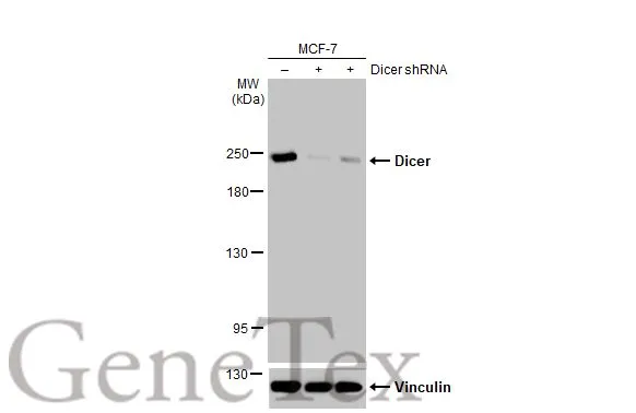
Non-transfected (–) and transfected (+) MCF-7 whole cell extracts (30 μg) were separated by 5% SDS-PAGE, and the membrane was blotted with Dicer antibody [HL1232] (GTX636580) diluted at 1:1000. The HRP-conjugated anti-rabbit IgG antibody (GTX213110-01) was used to detect the primary antibody.
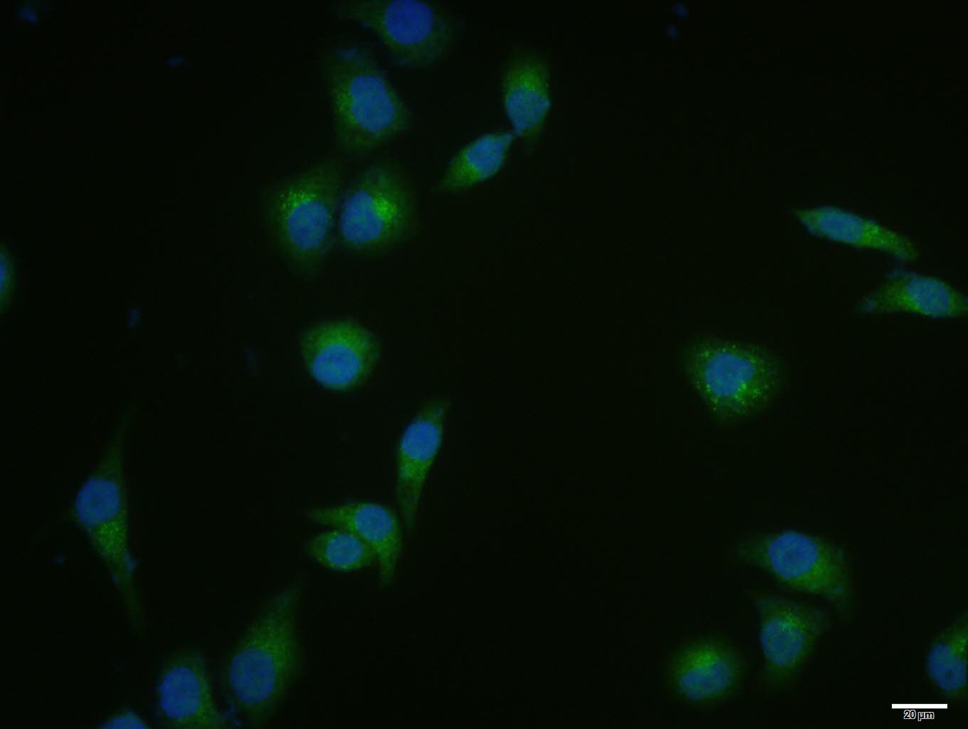
![Dicer antibody [HL1232] detects Dicer protein at cytoplasm by immunofluorescent analysis. Sample: HeLa cells were fixed in 4% paraformaldehyde at RT for 15 min. Green: Dicer stained by Dicer antibody [HL1232] (GTX636580) diluted at 1:500. Red: alpha Tubulin, a cytoskeleton marker, stained by alpha Tubulin antibody [GT114] (GTX628802) diluted at 1:1000. Blue: Fluoroshield with DAPI (GTX30920). Scale bar= 10μm.](https://www.genetex.com/upload/website/prouct_img/normal/GTX636580/GTX636580_T-44494_20220826_ICC_IF_22083119_510.webp "Dicer antibody [HL1232] detects Dicer protein at cytoplasm by immunofluorescent analysis. Sample: HeLa cells were fixed in 4% paraformaldehyde at RT for 15 min. Green: Dicer stained by Dicer antibody [HL1232] (GTX636580) diluted at 1:500. Red: alpha Tubulin, a cytoskeleton marker, stained by alpha Tubulin antibody [GT114] (GTX628802) diluted at 1:1000. Blue: Fluoroshield with DAPI (GTX30920). Scale bar= 10μm.")
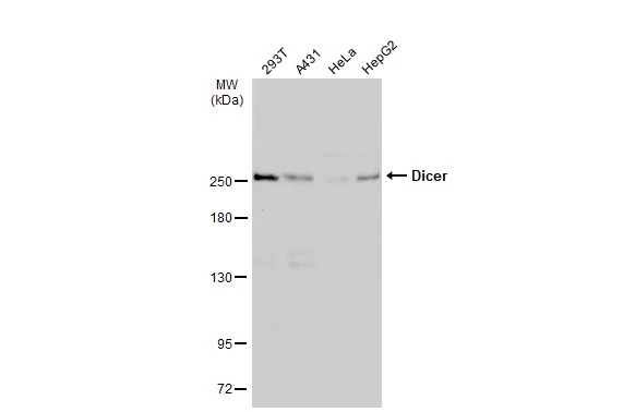
![Various whole cell extracts (30 μg) were separated by 5% SDS-PAGE, and the membrane was blotted with Dicer antibody [HL1232] (GTX636580) diluted at 1:1000. The HRP-conjugated anti-rabbit IgG antibody (GTX213110-01) was used to detect the primary antibody.](https://www.genetex.com/upload/website/prouct_img/normal/GTX636580/GTX636580_44536_20221209_WB_D_C_22121123_109.webp "Various whole cell extracts (30 μg) were separated by 5% SDS-PAGE, and the membrane was blotted with Dicer antibody [HL1232] (GTX636580) diluted at 1:1000. The HRP-conjugated anti-rabbit IgG antibody (GTX213110-01) was used to detect the primary antibody.")
![Various whole cell extracts (30 μg) were separated by 5% SDS-PAGE, and the membrane was blotted with Dicer antibody [HL1232] (GTX636580) diluted at 1:1000. The HRP-conjugated anti-rabbit IgG antibody (GTX213110-01) was used to detect the primary antibody.](https://www.genetex.com/upload/website/prouct_img/normal/GTX636580/GTX636580_44536_20211231_WB_w_23061202_729.webp "Various whole cell extracts (30 μg) were separated by 5% SDS-PAGE, and the membrane was blotted with Dicer antibody [HL1232] (GTX636580) diluted at 1:1000. The HRP-conjugated anti-rabbit IgG antibody (GTX213110-01) was used to detect the primary antibody.")
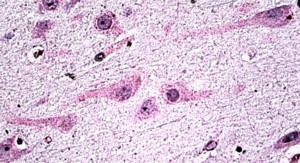
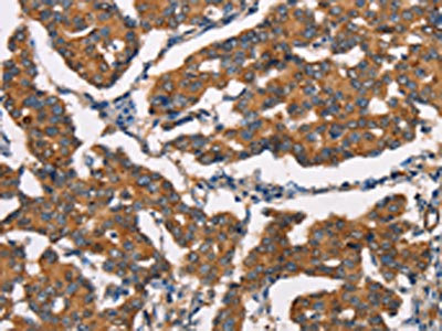
![Dicer antibody [HL1232] detects Dicer protein at cytoplasm by immunohistochemical analysis. Sample: Paraffin-embedded mouse testis. Dicer stained by Dicer antibody [HL1232] (GTX636580) diluted at 1:500. Antigen Retrieval: Citrate buffer, pH 6.0, 15 min](https://www.genetex.com/upload/website/prouct_img/normal/GTX636580/GTX636580_T-44494_20220114_IHC-P_M_w_23061202_859.webp "Dicer antibody [HL1232] detects Dicer protein at cytoplasm by immunohistochemical analysis. Sample: Paraffin-embedded mouse testis. Dicer stained by Dicer antibody [HL1232] (GTX636580) diluted at 1:500. Antigen Retrieval: Citrate buffer, pH 6.0, 15 min")
![Various whole cell extracts (30 μg) were separated by 5% SDS-PAGE, and the membrane was blotted with Dicer antibody [HL1232] (GTX636580) diluted at 1:1000. The HRP-conjugated anti-rabbit IgG antibody (GTX213110-01) was used to detect the primary antibody, and the signal was developed with Trident ECL plus-Enhanced.](https://www.genetex.com/upload/website/prouct_img/normal/GTX636580/GTX636580_T-44494_20211126_WB_R_w_23061202_994.webp "Various whole cell extracts (30 μg) were separated by 5% SDS-PAGE, and the membrane was blotted with Dicer antibody [HL1232] (GTX636580) diluted at 1:1000. The HRP-conjugated anti-rabbit IgG antibody (GTX213110-01) was used to detect the primary antibody, and the signal was developed with Trident ECL plus-Enhanced.")
![Various whole cell extracts (30 μg) were separated by 5% SDS-PAGE, and the membrane was blotted with Dicer antibody [HL1232] (GTX636580) diluted at 1:1000. The HRP-conjugated anti-rabbit IgG antibody (GTX213110-01) was used to detect the primary antibody, and the signal was developed with Trident ECL plus-Enhanced.](https://www.genetex.com/upload/website/prouct_img/normal/GTX636580/GTX636580_T-44494_20211126_WB_M_w_23061202_417.webp "Various whole cell extracts (30 μg) were separated by 5% SDS-PAGE, and the membrane was blotted with Dicer antibody [HL1232] (GTX636580) diluted at 1:1000. The HRP-conjugated anti-rabbit IgG antibody (GTX213110-01) was used to detect the primary antibody, and the signal was developed with Trident ECL plus-Enhanced.")
Non-transfected (–) and transfected (+) MCF-7 whole cell extracts (30 μg) were separated by 5% SDS-PAGE, and the membrane was blotted with Dicer antibody [HL1232] (GTX636580) diluted at 1:1000. The HRP-conjugated anti-rabbit IgG antibody (GTX213110-01) was used to detect the primary antibody.
Dicer antibody [HL1232]
GTX636580
ApplicationsImmunoFluorescence, Western Blot, ImmunoCytoChemistry, ImmunoHistoChemistry, ImmunoHistoChemistry Paraffin
Product group Antibodies
ReactivityCanine, Feline, Human, Mouse, Rat
TargetDICER1
Overview
- SupplierGeneTex
- Product NameDicer antibody [HL1232]
- Delivery Days Customer9
- Application Supplier NoteWB: 1:500-1:3000. *Optimal dilutions/concentrations should be determined by the researcher.Not tested in other applications.
- ApplicationsImmunoFluorescence, Western Blot, ImmunoCytoChemistry, ImmunoHistoChemistry, ImmunoHistoChemistry Paraffin
- CertificationResearch Use Only
- ClonalityMonoclonal
- Clone IDHL1232
- Concentration1 mg/ml
- ConjugateUnconjugated
- Gene ID23405
- Target nameDICER1
- Target descriptiondicer 1, ribonuclease III
- Target synonymsDCR1, Dicer, Dicer1e, GLOW, HERNA, K12H4.8-LIKE, MNG1, RMSE2, aviD, endoribonuclease Dicer, Dicer1, Dcr-1 homolog, dicer 1, double-stranded RNA-specific endoribonuclease, dicer 1, ribonuclease type III, helicase MOI, helicase with RNAse motif
- HostRabbit
- IsotypeIgG
- Protein IDQ9UPY3
- Protein NameEndoribonuclease Dicer
- Scientific DescriptionThis gene encodes a protein possessing an RNA helicase motif containing a DEXH box in its amino terminus and an RNA motif in the carboxy terminus. The encoded protein functions as a ribonuclease and is required by the RNA interference and small temporal RNA (stRNA) pathways to produce the active small RNA component that represses gene expression. Alternative splicing results in multiple transcript variants. [provided by RefSeq, Sep 2010]
- ReactivityCanine, Feline, Human, Mouse, Rat
- Storage Instruction-20°C or -80°C,2°C to 8°C
- UNSPSC41116161
Datasheet
Related products

Product group Antibodies
Anti-DICER1 AntibodyAMAB90737
ApplicationsWestern Blot, ImmunoCytoChemistry, ImmunoHistoChemistry
ReactivityHuman
TargetDICER1
- SizePrice
Product group Antibodies
Anti-Dicer AntibodyA326239
ApplicationsELISA, ImmunoHistoChemistry
ReactivityHuman
- SizePrice
Product group Antibodies
DICER1 AntibodyCSB-PA447308
ApplicationsELISA, ImmunoHistoChemistry
ReactivityHuman, Mouse
TargetDICER1
- SizePrice
Product group Antibodies
DICER1 Polyclonal AntibodyBS-6697R
ApplicationsImmunoFluorescence, Western Blot, ELISA, ImmunoCytoChemistry, ImmunoHistoChemistry, ImmunoHistoChemistry Frozen, ImmunoHistoChemistry Paraffin
ReactivityBovine, Equine, Human, Mouse, Porcine, Rat
TargetDICER1
- SizePrice

Product group Antibodies
Anti-Dicer/DICER1 Antibody Picoband(r)A00190-2-CARRIER-FREE
ApplicationsFlow Cytometry, ImmunoFluorescence, Western Blot, ELISA, ImmunoCytoChemistry
ReactivityHuman, Mouse
TargetDICER1
- SizePrice
Product group Antibodies

Goat anti-Dicer1EB06871
ApplicationsELISA, ImmunoHistoChemistry
ReactivityBovine, Canine, Human, Mouse, Porcine, Rat
TargetDICER1
- SizePrice
Product group Antibodies
ApplicationsImmunoPrecipitation, Western Blot, ImmunoCytoChemistry, ImmunoHistoChemistry
ReactivityMouse, Rat
TargetDICER1
- SizePrice
Product group Antibodies

DICER1 / Dicer AntibodyLS-C403112
ApplicationsELISA, ImmunoHistoChemistry
ReactivityHuman, Mouse
TargetDICER1
- SizePrice
Product group Antibodies
Dicer antibodyGTX130536
ApplicationsWestern Blot
ReactivityHuman, Mouse
TargetDICER1
- SizePrice
![Dicer antibody [GT1176] detects Dicer protein at cytoplasm by immunofluorescent analysis. Sample: A431 cells were fixed in 4% paraformaldehyde at RT for 15 min. Green: Dicer stained by Dicer antibody [GT1176] (GTX633917) diluted at 1:500. Blue: Hoechst 33342 staining. Scale bar= 10μm.](https://www.genetex.com/upload/website/prouct_img/normal/GTX633917/GTX633917_42814_20180321_ICC_IF_w_23061202_996.webp)
Product group Antibodies
Dicer antibody [GT1176]GTX633917
ApplicationsImmunoFluorescence, Western Blot, ImmunoCytoChemistry
ReactivityHuman
TargetDICER1
- SizePrice