

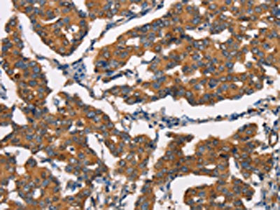
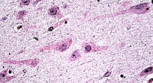
The image on the left is immunohistochemistry of paraffin-embedded Human breast cancer tissue using CSB-PA447308(DICER1 Antibody) at dilution 1/25, on the right is treated with synthetic peptide. (Original magnification: x200)
 at dilution 1/25, on the right is treated with synthetic peptide. (Original magnification: x200)")
The image on the left is immunohistochemistry of paraffin-embedded Human breast cancer tissue using CSB-PA447308(DICER1 Antibody) at dilution 1/25, on the right is treated with synthetic peptide. (Original magnification: x200)
DICER1 Antibody
CSB-PA447308
ApplicationsELISA, ImmunoHistoChemistry
Product group Antibodies
ReactivityHuman, Mouse
TargetDICER1
Overview
- SupplierCusabio
- Product NameDICER1 Antibody
- Delivery Days Customer20
- ApplicationsELISA, ImmunoHistoChemistry
- CertificationResearch Use Only
- ClonalityPolyclonal
- ConjugateUnconjugated
- Gene ID23405
- Target nameDICER1
- Target descriptiondicer 1, ribonuclease III
- Target synonymsDCR1, Dicer, Dicer1e, GLOW, HERNA, K12H4.8-LIKE, MNG1, RMSE2, aviD, endoribonuclease Dicer, Dicer1, Dcr-1 homolog, dicer 1, double-stranded RNA-specific endoribonuclease, dicer 1, ribonuclease type III, helicase MOI, helicase with RNAse motif
- HostRabbit
- IsotypeIgG
- Protein IDQ9UPY3
- Protein NameEndoribonuclease Dicer
- Scientific DescriptionThis gene encodes a protein possessing an RNA helicase motif containing a DEXH box in its amino terminus and an RNA motif in the carboxy terminus. The encoded protein functions as a ribonuclease and is required by the RNA interference and small temporal RNA (stRNA) pathways to produce the active small RNA component that represses gene expression. Alternative splicing results in multiple transcript variants.
- ReactivityHuman, Mouse
- Storage Instruction-20°C or -80°C
- UNSPSC41116161
Related products

Product group Antibodies
Anti-Dicer/DICER1 Antibody Picoband(r)A00190-2-CARRIER-FREE
ApplicationsFlow Cytometry, ImmunoFluorescence, Western Blot, ELISA, ImmunoCytoChemistry
ReactivityHuman, Mouse
TargetDICER1
- SizePrice

Product group Antibodies
Anti-DICER1 AntibodyAMAB90737
ApplicationsWestern Blot, ImmunoCytoChemistry, ImmunoHistoChemistry
ReactivityHuman
TargetDICER1
- SizePrice
Product group Antibodies
Anti-Dicer AntibodyA326239
ApplicationsELISA, ImmunoHistoChemistry
ReactivityHuman
- SizePrice
Product group Antibodies

Goat anti-Dicer1EB06871
ApplicationsELISA, ImmunoHistoChemistry
ReactivityBovine, Canine, Human, Mouse, Porcine, Rat
TargetDICER1
- SizePrice
Product group Antibodies

DICER1 / Dicer AntibodyLS-C403112
ApplicationsELISA, ImmunoHistoChemistry
ReactivityHuman, Mouse
TargetDICER1
- SizePrice
Product group Antibodies
ApplicationsImmunoPrecipitation, Western Blot, ImmunoCytoChemistry, ImmunoHistoChemistry
ReactivityMouse, Rat
TargetDICER1
- SizePrice
Product group Antibodies
Dicer antibodyGTX130536
ApplicationsWestern Blot
ReactivityHuman, Mouse
TargetDICER1
- SizePrice
Product group Antibodies
DICER1 Polyclonal AntibodyBS-6697R
ApplicationsImmunoFluorescence, Western Blot, ELISA, ImmunoCytoChemistry, ImmunoHistoChemistry, ImmunoHistoChemistry Frozen, ImmunoHistoChemistry Paraffin
ReactivityBovine, Equine, Human, Mouse, Porcine, Rat
TargetDICER1
- SizePrice