

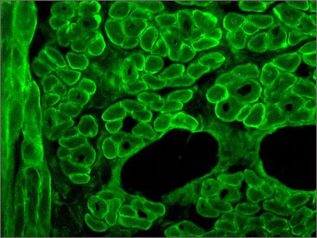
IHC-Fr analysis of acetone-fixed human tongue tissue using GTX27163 Dystrophin antibody [MANDYS8] at 1:400.
![IHC-Fr analysis of acetone-fixed human tongue tissue using GTX27163 Dystrophin antibody [MANDYS8] at 1:400.](https://www.genetex.com/upload/website/prouct_img/normal/GTX27163/GTX27163_20170605_IHC-Fr_1_w_23060722_664.webp "IHC-Fr analysis of acetone-fixed human tongue tissue using GTX27163 Dystrophin antibody [MANDYS8] at 1:400.")
IHC-Fr analysis of acetone-fixed human tongue tissue using GTX27163 Dystrophin antibody [MANDYS8] at 1:400.
Dystrophin antibody [MANDYS8]
GTX27163
ApplicationsImmunoFluorescence, Western Blot, ELISA, ImmunoCytoChemistry, ImmunoHistoChemistry, ImmunoHistoChemistry Frozen
Product group Antibodies
ReactivityChicken, Human, Mouse, Porcine, Rabbit, Rat
TargetDMD
Overview
- SupplierGeneTex
- Product NameDystrophin antibody [MANDYS8]
- Delivery Days Customer9
- Application Supplier NoteIHC-Fr: 1:400. *Optimal dilutions/concentrations should be determined by the researcher.Not tested in other applications.
- ApplicationsImmunoFluorescence, Western Blot, ELISA, ImmunoCytoChemistry, ImmunoHistoChemistry, ImmunoHistoChemistry Frozen
- CertificationResearch Use Only
- ClonalityMonoclonal
- Clone IDMANDYS8
- ConjugateUnconjugated
- Gene ID1756
- Target nameDMD
- Target descriptiondystrophin
- Target synonymsBMD, CMD3B, DXS142, DXS164, DXS206, DXS230, DXS239, DXS268, DXS269, DXS270, DXS272, MRX85, dystrophin, mutant dystrophin
- HostMouse
- IsotypeIgG2b
- Protein IDP11532
- Protein NameDystrophin
- Scientific DescriptionThe rod domain of the human dystrophin molecule is present in normal muscle tissue and in nearly all Becker muscular dystrophies. It is absent in the cases of Duchenne muscular dystrophies and in the dystrophic mouse (mdx).
- ReactivityChicken, Human, Mouse, Porcine, Rabbit, Rat
- Storage Instruction-20°C or -80°C,2°C to 8°C
- UNSPSC41116161
References
- A dystrophic Duchenne mouse model for testing human antisense oligonucleotides. Veltrop M et al., 2018, PLoS OneRead this paper
Datasheet
Related products
Product group Antibodies

Anti-Dystrophin [133D7-1], Human IgG1, kappaAB04679-10.0
ReactivityHuman
TargetDMD
- SizePrice
Product group Antibodies
Anti-Dystrophin AntibodyA17013
ApplicationsImmunoFluorescence, Western Blot, ImmunoCytoChemistry, ImmunoHistoChemistry
ReactivityHuman, Mouse, Rat
- SizePrice
Product group Antibodies
Anti-DMD Antibody144-63081
ApplicationsImmunoFluorescence, Western Blot, ImmunoHistoChemistry
ReactivityHuman, Mouse, Rat
TargetDMD
- SizePrice
Product group Antibodies

DMD / Dystrophin Antibody (Biotin)LS-C680389
ApplicationsELISA
ReactivityHuman
TargetDMD
- SizePrice
Product group Antibodies
Dystrophin Recombinant AntibodyBSM-61024R
ApplicationsImmunoFluorescence, ImmunoHistoChemistry, ImmunoHistoChemistry Frozen, ImmunoHistoChemistry Paraffin
ReactivityHuman, Mouse, Rat
TargetDMD
- SizePrice
Product group Antibodies
DMD AntibodyCSB-PA006963LA01HU
ApplicationsELISA, ImmunoHistoChemistry
ReactivityHuman
TargetDMD
- SizePrice
Product group Antibodies
ApplicationsImmunoPrecipitation, Western Blot, ImmunoCytoChemistry, ImmunoHistoChemistry
ReactivityMouse, Porcine, Rat
TargetDMD
- SizePrice
Product group Antibodies
Dystrophin antibody [Dy8/6C5]GTX01868
ApplicationsImmunoHistoChemistry, ImmunoHistoChemistry Frozen
ReactivityCanine, Chicken, Hamster, Human, Mouse, Rabbit, Rat
TargetDMD
- SizePrice
Product group Antibodies
Dystrophin antibody [Dy10/12B2]GTX01869
ApplicationsImmunoHistoChemistry, ImmunoHistoChemistry Frozen
ReactivityHuman
TargetDMD
- SizePrice