

Dystrophin Recombinant Antibody
BSM-61024R
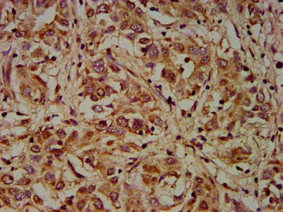
ApplicationsImmunoFluorescence, ImmunoHistoChemistry, ImmunoHistoChemistry Frozen, ImmunoHistoChemistry Paraffin
Product group Antibodies
ReactivityHuman, Mouse, Rat
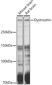
TargetDMD
Overview
- SupplierBioss
- Product NameDystrophin Recombinant Antibody
- Delivery Days Customer16
- ApplicationsImmunoFluorescence, ImmunoHistoChemistry, ImmunoHistoChemistry Frozen, ImmunoHistoChemistry Paraffin
- Applications SupplierIHC-P(1:200-400), IHC-F(1:100-500), IF()
- CertificationResearch Use Only
- ClonalityMonoclonal
- ConjugateUnconjugated
- Gene ID1756
- Target nameDMD
- Target descriptiondystrophin
- Target synonymsBMD, CMD3B, DXS142, DXS164, DXS206, DXS230, DXS239, DXS268, DXS269, DXS270, DXS272, MRX85, dystrophin, mutant dystrophin
- HostRabbit
- IsotypeIgG
- Protein IDP11532
- Protein NameDystrophin
- ReactivityHuman, Mouse, Rat
- Storage Instruction-20°C,2°C to 8°C
- UNSPSC41116161
Related products
Product group Antibodies

Anti-Dystrophin [133D7-1], Human IgG1, kappaAB04679-10.0
ReactivityHuman
TargetDMD
- SizePrice
Product group Antibodies
Anti-Dystrophin AntibodyA17013
ApplicationsImmunoFluorescence, Western Blot, ImmunoCytoChemistry, ImmunoHistoChemistry
ReactivityHuman, Mouse, Rat
- SizePrice
Product group Antibodies
Anti-DMD Antibody144-63081
ApplicationsImmunoFluorescence, Western Blot, ImmunoHistoChemistry
ReactivityHuman, Mouse, Rat
TargetDMD
- SizePrice
Product group Antibodies

DMD / Dystrophin Antibody (Biotin)LS-C680389
ApplicationsELISA
ReactivityHuman
TargetDMD
- SizePrice
Product group Antibodies
DMD AntibodyCSB-PA006963LA01HU
ApplicationsELISA, ImmunoHistoChemistry
ReactivityHuman
TargetDMD
- SizePrice
Product group Antibodies
ApplicationsImmunoPrecipitation, Western Blot, ImmunoCytoChemistry, ImmunoHistoChemistry
ReactivityMouse, Porcine, Rat
TargetDMD
- SizePrice
Product group Antibodies
Dystrophin antibody [Dy8/6C5]GTX01868
ApplicationsImmunoHistoChemistry, ImmunoHistoChemistry Frozen
ReactivityCanine, Chicken, Hamster, Human, Mouse, Rabbit, Rat
TargetDMD
- SizePrice

Product group Antibodies
Anti-DMD AntibodyHPA002725
ApplicationsImmunoHistoChemistry
ReactivityHuman
TargetDMD
- SizePrice