

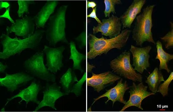
eIF4E antibody [HL1553] detects eIF4E protein at cytoplasm by immunofluorescent analysis. Sample: HeLa cells were fixed in 4% paraformaldehyde at RT for 15 min. Green: eIF4E stained by eIF4E antibody [HL1553] (GTX637028) diluted at 1:500. Red: alpha Tubulin, a cytoskeleton marker, stained by alpha Tubulin antibody [GT114] (GTX628802) diluted at 1:1000. Blue: Fluoroshield with DAPI (GTX30920).
![Mouse tissue extract (50 μg) was separated by 12% SDS-PAGE, and the membrane was blotted with eIF4E antibody [HL1553] (GTX637028) diluted at 1:1000. The HRP-conjugated anti-rabbit IgG antibody (GTX213110-01) was used to detect the primary antibody.](https://www.genetex.com/upload/website/prouct_img/normal/GTX637028/GTX637028_T-44697_20220715_WB_M_testis_22071823_262.webp "Mouse tissue extract (50 μg) was separated by 12% SDS-PAGE, and the membrane was blotted with eIF4E antibody [HL1553] (GTX637028) diluted at 1:1000. The HRP-conjugated anti-rabbit IgG antibody (GTX213110-01) was used to detect the primary antibody.")
![Various whole cell extracts (30 μg) were separated by 12% SDS-PAGE, and the membrane was blotted with eIF4E antibody [HL1553] (GTX637028) diluted at 1:1000. The HRP-conjugated anti-rabbit IgG antibody (GTX213110-01) was used to detect the primary antibody.](https://www.genetex.com/upload/website/prouct_img/normal/GTX637028/GTX637028_T-44697_20220715_WB_M_R_22071823_579.webp "Various whole cell extracts (30 μg) were separated by 12% SDS-PAGE, and the membrane was blotted with eIF4E antibody [HL1553] (GTX637028) diluted at 1:1000. The HRP-conjugated anti-rabbit IgG antibody (GTX213110-01) was used to detect the primary antibody.")
![eIF4E antibody [HL1553] detects eIF4E protein at cytoplasm by immunohistochemical analysis. Sample: Paraffin-embedded mouse kidney. eIF4E stained by eIF4E antibody [HL1553] (GTX637028) diluted at 1:100. Antigen Retrieval: Tris-EDTA buffer, pH 8.0, 15 min](https://www.genetex.com/upload/website/prouct_img/normal/GTX637028/GTX637028_T-44697_20220729_IHC-P_M_22081423_667.webp "eIF4E antibody [HL1553] detects eIF4E protein at cytoplasm by immunohistochemical analysis. Sample: Paraffin-embedded mouse kidney. eIF4E stained by eIF4E antibody [HL1553] (GTX637028) diluted at 1:100. Antigen Retrieval: Tris-EDTA buffer, pH 8.0, 15 min")
![Various whole cell extracts (30 μg) were separated by 12% SDS-PAGE, and the membrane was blotted with eIF4E antibody [HL1553] (GTX637028) diluted at 1:1000. The HRP-conjugated anti-rabbit IgG antibody (GTX213110-01) was used to detect the primary antibody.](https://www.genetex.com/upload/website/prouct_img/normal/GTX637028/GTX637028_44781_20220826_WB_22083119_286.webp "Various whole cell extracts (30 μg) were separated by 12% SDS-PAGE, and the membrane was blotted with eIF4E antibody [HL1553] (GTX637028) diluted at 1:1000. The HRP-conjugated anti-rabbit IgG antibody (GTX213110-01) was used to detect the primary antibody.")
![eIF4E antibody [HL1553] detects eIF4E protein by immunohistochemical analysis. Sample: Paraffin-embedded rat tissues. eIF4E stained by eIF4E antibody [HL1553] (GTX637028) diluted at 1:100. Antigen Retrieval: Citrate buffer, pH 6.0, 15 min](https://www.genetex.com/upload/website/prouct_img/normal/GTX637028/GTX637028_44781_20221227_IHC-P_multiple_R_22122821_181.webp "eIF4E antibody [HL1553] detects eIF4E protein by immunohistochemical analysis. Sample: Paraffin-embedded rat tissues. eIF4E stained by eIF4E antibody [HL1553] (GTX637028) diluted at 1:100. Antigen Retrieval: Citrate buffer, pH 6.0, 15 min")
![293T whole cell and cytoplasm extracts (30 μg) were separated by 15% SDS-PAGE, and the membrane was blotted with eIF4E antibody [HL1553] (GTX637028) diluted at 1:1000. The HRP-conjugated anti-rabbit IgG antibody (GTX213110-01) was used to detect the primary antibody.](https://www.genetex.com/upload/website/prouct_img/normal/GTX637028/GTX637028_44781_20230728_WB_Fraction_23073119_837.webp "293T whole cell and cytoplasm extracts (30 μg) were separated by 15% SDS-PAGE, and the membrane was blotted with eIF4E antibody [HL1553] (GTX637028) diluted at 1:1000. The HRP-conjugated anti-rabbit IgG antibody (GTX213110-01) was used to detect the primary antibody.")
eIF4E antibody [HL1553] detects eIF4E protein at cytoplasm by immunofluorescent analysis. Sample: HeLa cells were fixed in 4% paraformaldehyde at RT for 15 min. Green: eIF4E stained by eIF4E antibody [HL1553] (GTX637028) diluted at 1:500. Red: alpha Tubulin, a cytoskeleton marker, stained by alpha Tubulin antibody [GT114] (GTX628802) diluted at 1:1000. Blue: Fluoroshield with DAPI (GTX30920).
eIF4E antibody [HL1553]
GTX637028
ApplicationsImmunoFluorescence, Western Blot, ImmunoCytoChemistry, ImmunoHistoChemistry, ImmunoHistoChemistry Paraffin
Product group Antibodies
ReactivityHuman, Mouse, Rat
TargetEIF4E
Overview
- SupplierGeneTex
- Product NameeIF4E antibody [HL1553]
- Delivery Days Customer9
- Application Supplier NoteICC/IF: 1:100-1:1000. *Optimal dilutions/concentrations should be determined by the researcher.Not tested in other applications.
- ApplicationsImmunoFluorescence, Western Blot, ImmunoCytoChemistry, ImmunoHistoChemistry, ImmunoHistoChemistry Paraffin
- CertificationResearch Use Only
- ClonalityMonoclonal
- Clone IDHL1553
- Concentration1 mg/ml
- ConjugateUnconjugated
- Gene ID1977
- Target nameEIF4E
- Target descriptioneukaryotic translation initiation factor 4E
- Target synonymsAUTS19, CBP, EIF4E1, EIF4EL1, EIF4F, eIF-4E, eukaryotic translation initiation factor 4E, eIF-4F 25 kDa subunit, eukaryotic translation initiation factor 4E-like 1, mRNA cap-binding protein
- HostRabbit
- IsotypeIgG
- Protein IDP06730
- Protein NameEukaryotic translation initiation factor 4E
- Scientific DescriptionThe protein encoded by this gene is a component of the eukaryotic translation initiation factor 4F complex, which recognizes the 7-methylguanosine cap structure at the 5 end of messenger RNAs. The encoded protein aids in translation initiation by recruiting ribosomes to the 5-cap structure. Association of this protein with the 4F complex is the rate-limiting step in translation initiation. This gene acts as a proto-oncogene, and its expression and activation is associated with transformation and tumorigenesis. Several pseudogenes of this gene are found on other chromosomes. Alternative splicing results in multiple transcript variants. [provided by RefSeq, Sep 2015]
- ReactivityHuman, Mouse, Rat
- Storage Instruction-20°C or -80°C,2°C to 8°C
- UNSPSC12352203
Datasheet
Related products

Product group Antibodies
Anti-EIF4E Antibody144-60024
ApplicationsImmunoFluorescence, Western Blot
ReactivityHuman
TargetEIF4E
- SizePrice

Product group Antibodies
ApplicationsFlow Cytometry, ImmunoPrecipitation, Western Blot, ImmunoHistoChemistry
ReactivityHuman, Mouse, Rat
TargetEIF4E
- SizePrice
Product group Antibodies
References
eIF4E antibodyGTX132092
ApplicationsImmunoFluorescence, Western Blot, ImmunoCytoChemistry, ImmunoHistoChemistry, ImmunoHistoChemistry Paraffin
ReactivityHuman, Mouse
TargetEIF4E
- SizePrice
Product group Antibodies
eIF4E (phospho Ser209) antibodyGTX133606
ApplicationsWestern Blot
ReactivityHuman, Mouse
TargetEIF4E
- SizePrice
![WB analysis of HeLa (1), HEK293 (2) and K562 (3) cell lysate using GTX60420 eIF4E antibody [5D11].](https://www.genetex.com/upload/website/prouct_img/normal/GTX60420/GTX60420_20170912_WB_w_23061123_620.webp)
Product group Antibodies
eIF4E antibody [5D11]GTX60420
ApplicationsFlow Cytometry, ImmunoFluorescence, Western Blot, ELISA, ImmunoCytoChemistry, ImmunoHistoChemistry, ImmunoHistoChemistry Paraffin
ReactivityHuman
TargetEIF4E
- SizePrice
![Various whole cell extracts (30 μg) were separated by 12% SDS-PAGE, and the membrane was blotted with eIF4E antibody [HL1554] (GTX637029) diluted at 1:1000. The HRP-conjugated anti-rabbit IgG antibody (GTX213110-01) was used to detect the primary antibody.](https://www.genetex.com/upload/website/prouct_img/normal/GTX637029/GTX637029_T-44697_20220617_WB_M_R_22062121_206.webp)
Product group Antibodies
eIF4E antibody [HL1554]GTX637029
ApplicationsWestern Blot, ImmunoHistoChemistry, ImmunoHistoChemistry Paraffin
ReactivityHuman, Mouse, Rat
TargetEIF4E
- SizePrice
![Various whole cell extracts (30 μg) were separated by 12% SDS-PAGE, and the membrane was blotted with eIF4E antibody [HL1555] (GTX637030) diluted at 1:1000. The HRP-conjugated anti-rabbit IgG antibody (GTX213110-01) was used to detect the primary antibody.](https://www.genetex.com/upload/website/prouct_img/normal/GTX637030/GTX637030_T-44697_20220617_WB_M_R_22062121_522.webp)
Product group Antibodies
eIF4E antibody [HL1555]GTX637030
ApplicationsWestern Blot, ImmunoHistoChemistry, ImmunoHistoChemistry Paraffin
ReactivityHuman, Mouse, Rat
TargetEIF4E
- SizePrice
Product group Antibodies
Anti-EIF4EY058779
ApplicationsWestern Blot, ELISA, ImmunoHistoChemistry
ReactivityHuman, Mouse, Rat
- SizePrice
Product group Antibodies
References
eIF4E antibodyGTX82525
ApplicationsFlow Cytometry, Western Blot, ImmunoHistoChemistry, ImmunoHistoChemistry Paraffin
ReactivityHuman, Mammals
TargetEIF4E
- SizePrice