

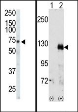
(LEFT)Western blot analysis of anti-EphA3 Pab in CHO cell lysate. EphA3 (arrow) was detected using purified Pab. Secondary HRP-anti-rabbit was used for signal visualization with chemiluminescence. (RIGHT)Western blot analysis of EphA3 (arrow) using rabbit polyclonal EphA3 Antibody (C-term). 293 cell lysates (2 ug/lane) either nontransfected (Lane 1) or transiently transfected with the EphA3 gene (Lane 2)
(LEFT)Western blot analysis of anti-EphA3 Pab in CHO cell lysate. EphA3 (arrow) was detected using purified Pab. Secondary HRP-anti-rabbit was used for signal visualization with chemiluminescence. (RIGHT)Western blot analysis of EphA3 (arrow) using rabbit polyclonal EphA3 Antibody (C-term). 293 cell lysates (2 ug/lane) either nontransfected (Lane 1) or transiently transfected with the EphA3 gene (Lane 2)
Eph receptor A3 (EPHA3) (C-term) Rabbit Polyclonal Antibody
AP14277PU-N
ApplicationsWestern Blot, ImmunoHistoChemistry
Product group Antibodies
ReactivityHamster, Human
TargetEPHA3
Overview
- SupplierOriGene
- Product NameEph receptor A3 (EPHA3) (C-term) Rabbit Polyclonal Antibody
- Delivery Days Customer14
- ApplicationsWestern Blot, ImmunoHistoChemistry
- CertificationResearch Use Only
- ClonalityPolyclonal
- Gene ID2042
- Target nameEPHA3
- Target descriptionEPH receptor A3
- Target synonymsEK4, ETK, ETK1, HEK, HEK4, TYRO4, ephrin type-A receptor 3, EPH-like kinase 4, TYRO4 protein tyrosine kinase, eph-like tyrosine kinase 1, human embryo kinase 1, testicular tissue protein Li 64, tyrosine-protein kinase receptor ETK1
- HostRabbit
- Protein IDP29320
- Protein NameEphrin type-A receptor 3
- Scientific DescriptionEph receptor A3 (EPHA3) (C-term) rabbit polyclonal antibody, Purified
- ReactivityHamster, Human
- Storage Instruction-20°C,2°C to 8°C
- UNSPSC12352203
MSDS
Related products

Product group Antibodies
Anti-Eph receptor A3/EPHA3 Antibody Picoband(r)A02872-1-CARRIER-FREE
ApplicationsFlow Cytometry, ImmunoFluorescence, Western Blot, ELISA, ImmunoCytoChemistry, ImmunoHistoChemistry
ReactivityHuman
TargetEPHA3
- SizePrice
Product group Antibodies

Anti-EphA3 [KB004]Ab02488-1.1
ApplicationsFunctional Assay
ReactivityHuman
TargetEPHA3
- SizePrice
Product group Antibodies
Anti-EPHA3 Antibody144-08414
ApplicationsWestern Blot, ImmunoHistoChemistry
ReactivityHuman, Mouse, Rat
TargetEPHA3
- SizePrice
Product group Antibodies
EphA3 Polyclonal AntibodyBS-7032R
ApplicationsImmunoFluorescence, Western Blot, ELISA, ImmunoCytoChemistry, ImmunoHistoChemistry, ImmunoHistoChemistry Frozen, ImmunoHistoChemistry Paraffin
ReactivityBovine, Canine, Equine, Human, Mouse, Rat, Sheep
TargetEPHA3
- SizePrice
Product group Antibodies
EPHA3 AntibodyCSB-PA008057
ApplicationsWestern Blot, ELISA
ReactivityHuman, Mouse, Rat
TargetEPHA3
- SizePrice

Product group Antibodies
Anti-EPHA3 AntibodyHPA069390
ApplicationsImmunoCytoChemistry
ReactivityHuman
TargetEPHA3
- SizePrice
![Whole cell extract (30 μg) was separated by 7.5% SDS-PAGE, and the membrane was blotted with EphA3 antibody [N1N3] (GTX114067) diluted at 1:1500. The HRP-conjugated anti-rabbit IgG antibody (GTX213110-01) was used to detect the primary antibody, and the signal was developed with Trident ECL plus-Enhanced.](https://www.genetex.com/upload/website/prouct_img/normal/GTX114067/GTX114067_40660_20211015_WB_M_w_23060501_869.webp)
Product group Antibodies
EphA3 antibody [N1N3]GTX114067
ApplicationsWestern Blot, ImmunoHistoChemistry, ImmunoHistoChemistry Paraffin
ReactivityHuman, Mouse
TargetEPHA3
- SizePrice
Product group Antibodies
Anti-EPHA3 AntibodyA97563
ApplicationsWestern Blot, ELISA
ReactivityHuman, Mouse, Rat
- SizePrice