

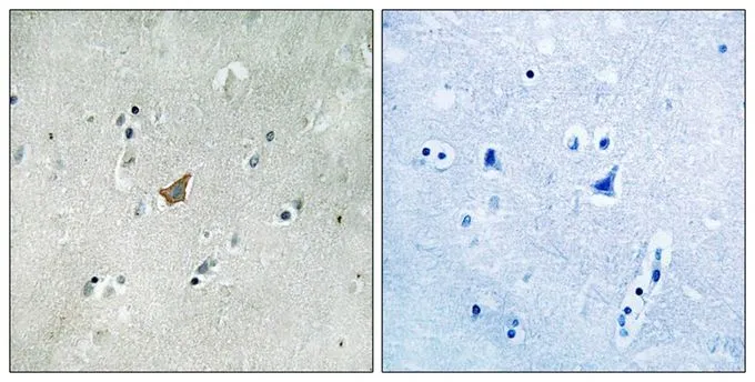
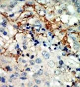
IHC-P analysis of human brain tissue using GTX17348 EphA3 (phospho Tyr779) / EphA4 (phospho Tyr779) / EphA5 (phospho Tyr833) antibody. Left : Primary antibody Right : Primary antibody pre-incubated with the antigen specific peptide
IHC-P analysis of human brain tissue using GTX17348 EphA3 (phospho Tyr779) / EphA4 (phospho Tyr779) / EphA5 (phospho Tyr833) antibody. Left : Primary antibody Right : Primary antibody pre-incubated with the antigen specific peptide
EphA3 (phospho Tyr779) / EphA4 (phospho Tyr779) / EphA5 (phospho Tyr833) antibody
GTX17348
ApplicationsImmunoHistoChemistry, ImmunoHistoChemistry Frozen, ImmunoHistoChemistry Paraffin
Product group Antibodies
ReactivityHuman
TargetEPHA3
Overview
- SupplierGeneTex
- Product NameEphA3 (phospho Tyr779) / EphA4 (phospho Tyr779) / EphA5 (phospho Tyr833) antibody
- Delivery Days Customer9
- Application Supplier NoteIHC-P: 1:50-1:100. *Optimal dilutions/concentrations should be determined by the researcher.Not tested in other applications.
- ApplicationsImmunoHistoChemistry, ImmunoHistoChemistry Frozen, ImmunoHistoChemistry Paraffin
- CertificationResearch Use Only
- ClonalityPolyclonal
- Concentration1 mg/ml
- ConjugateUnconjugated
- Gene ID2042
- Target nameEPHA3
- Target descriptionEPH receptor A3
- Target synonymsEK4, ETK, ETK1, HEK, HEK4, TYRO4, ephrin type-A receptor 3, EPH-like kinase 4, TYRO4 protein tyrosine kinase, eph-like tyrosine kinase 1, human embryo kinase 1, testicular tissue protein Li 64, tyrosine-protein kinase receptor ETK1
- HostRabbit
- IsotypeIgG
- Protein IDP29320
- Protein NameEphrin type-A receptor 3
- ReactivityHuman
- Storage Instruction-20°C or -80°C,2°C to 8°C
- UNSPSC41116161
References
- Identification and Therapeutic Intervention of Coactivated Anaplastic Lymphoma Kinase, Fibroblast Growth Factor Receptor 2, and Ephrin Type-A Receptor 5 Kinases in Hepatocellular Carcinoma. Wang X et al., 2019 Feb, HepatologyRead this paper
Datasheet
Related products
Product group Antibodies
Anti-EPHA3 AntibodyA97563
ApplicationsWestern Blot, ELISA
ReactivityHuman, Mouse, Rat
- SizePrice
Product group Antibodies

Anti-EphA3 [KB004]Ab02488-1.1
ApplicationsFunctional Assay
ReactivityHuman
TargetEPHA3
- SizePrice

Product group Antibodies
Anti-Eph receptor A3/EPHA3 Antibody Picoband(r)A02872-1-CARRIER-FREE
ApplicationsFlow Cytometry, ImmunoFluorescence, Western Blot, ELISA, ImmunoCytoChemistry, ImmunoHistoChemistry
ReactivityHuman
TargetEPHA3
- SizePrice
Product group Antibodies
Anti-EPHA3 Antibody144-08414
ApplicationsWestern Blot, ImmunoHistoChemistry
ReactivityHuman, Mouse, Rat
TargetEPHA3
- SizePrice
Product group Antibodies

EPHA3 / EPH Receptor A3 AntibodyLS-C831062
ApplicationsWestern Blot, ELISA
ReactivityHuman, Mouse, Rat
TargetEPHA3
- SizePrice
Product group Antibodies
EphA3 Polyclonal AntibodyBS-7032R
ApplicationsImmunoFluorescence, Western Blot, ELISA, ImmunoCytoChemistry, ImmunoHistoChemistry, ImmunoHistoChemistry Frozen, ImmunoHistoChemistry Paraffin
ReactivityBovine, Canine, Equine, Human, Mouse, Rat, Sheep
TargetEPHA3
- SizePrice
Product group Antibodies
EPHA3 AntibodyCSB-PA008057
ApplicationsWestern Blot, ELISA
ReactivityHuman, Mouse, Rat
TargetEPHA3
- SizePrice
Product group Antibodies
EphA3 antibody, N-termGTX81361
ApplicationsWestern Blot, ImmunoHistoChemistry, ImmunoHistoChemistry Paraffin
ReactivityHuman
TargetEPHA3
- SizePrice
![WB analysis of truncated Trx-EphA3 recombinant protein (1) andtruncated EphA3(aa566-983)-hIgGFc transfected CHO-K1 cell lysate(2) using GTX83180 EphA3 antibody [6C1B6].](https://www.genetex.com/upload/website/prouct_img/normal/GTX83180/GTX83180_20170912_WB_w_23061322_228.webp)
Product group Antibodies
EphA3 antibody [6C1B6]GTX83180
ApplicationsWestern Blot, ELISA
ReactivityHuman
TargetEPHA3
- SizePrice

Product group Antibodies
Anti-EPHA3 AntibodyHPA069390
ApplicationsImmunoCytoChemistry
ReactivityHuman
TargetEPHA3
- SizePrice