

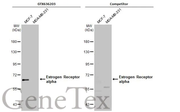
Various whole cell extracts (30 μg) were separated by 7.5% SDS-PAGE, and the membranes were blotted with Estrogen Receptor alpha antibody [HL1082] (GTX636203) diluted at 1:1000 and competitor's antibody (Competitor) diluted at 1:100. The HRP-conjugated anti-rabbit IgG antibody (GTX213110-01) was used to detect the primary antibody, and the signal was developed with Trident ECL plus-Enhanced. *The competitor is not affiliated with GeneTex and does not endorse this product.
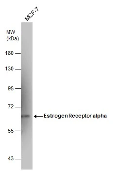
![Non-transfected (–) and transfected (+) 293T whole cell extracts (30 μg) were separated by 7.5% SDS-PAGE, and the membrane was blotted with Estrogen Receptor alpha antibody [HL1082] (GTX636203) diluted at 1:50000. The HRP-conjugated anti-rabbit IgG antibody (GTX213110-01) was used to detect the primary antibody.](https://www.genetex.com/upload/website/prouct_img/normal/GTX636203/GTX636203_44382_20210730_WB_B_w_23061202_936.webp "Non-transfected (–) and transfected (+) 293T whole cell extracts (30 μg) were separated by 7.5% SDS-PAGE, and the membrane was blotted with Estrogen Receptor alpha antibody [HL1082] (GTX636203) diluted at 1:50000. The HRP-conjugated anti-rabbit IgG antibody (GTX213110-01) was used to detect the primary antibody.")
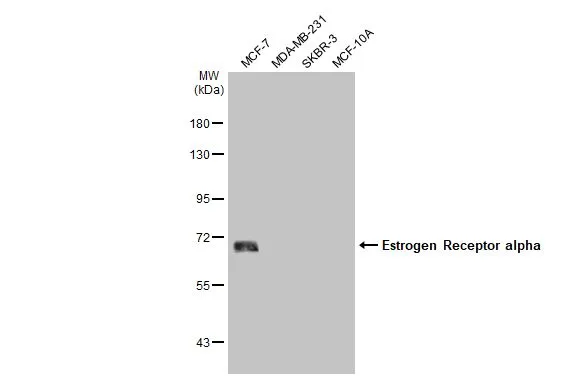
![Various whole cell extracts (30 μg) were separated by 7.5% SDS-PAGE, and the membrane was blotted with Estrogen Receptor alpha antibody [HL1082] (GTX636203) diluted at 1:1000. The HRP-conjugated anti-rabbit IgG antibody (GTX213110-01) was used to detect the primary antibody.](https://www.genetex.com/upload/website/prouct_img/normal/GTX636203/GTX636203_44382_20210723_WB_w_23061202_713.webp "Various whole cell extracts (30 μg) were separated by 7.5% SDS-PAGE, and the membrane was blotted with Estrogen Receptor alpha antibody [HL1082] (GTX636203) diluted at 1:1000. The HRP-conjugated anti-rabbit IgG antibody (GTX213110-01) was used to detect the primary antibody.")
![Estrogen Receptor alpha antibody [HL1082] detects Estrogen Receptor alpha protein at nucleus by immunohistochemical analysis. Sample: Paraffin-embedded human breast carcinoma. Estrogen Receptor alpha stained by Estrogen Receptor alpha antibody [HL1082] (GTX636203) diluted at 1:100. Antigen Retrieval: Citrate buffer, pH 6.0, 15 min](https://www.genetex.com/upload/website/prouct_img/normal/GTX636203/GTX636203_44382_20210813_IHC-P_w_23061202_287.webp "Estrogen Receptor alpha antibody [HL1082] detects Estrogen Receptor alpha protein at nucleus by immunohistochemical analysis. Sample: Paraffin-embedded human breast carcinoma. Estrogen Receptor alpha stained by Estrogen Receptor alpha antibody [HL1082] (GTX636203) diluted at 1:100. Antigen Retrieval: Citrate buffer, pH 6.0, 15 min")
Various whole cell extracts (30 μg) were separated by 7.5% SDS-PAGE, and the membranes were blotted with Estrogen Receptor alpha antibody [HL1082] (GTX636203) diluted at 1:1000 and competitor's antibody (Competitor) diluted at 1:100. The HRP-conjugated anti-rabbit IgG antibody (GTX213110-01) was used to detect the primary antibody, and the signal was developed with Trident ECL plus-Enhanced. *The competitor is not affiliated with GeneTex and does not endorse this product.
Estrogen Receptor alpha antibody [HL1082]
GTX636203
ApplicationsWestern Blot, ImmunoHistoChemistry, ImmunoHistoChemistry Paraffin
Product group Antibodies
ReactivityHuman
TargetESR1
Overview
- SupplierGeneTex
- Product NameEstrogen Receptor alpha antibody [HL1082]
- Delivery Days Customer9
- Application Supplier NoteWB: 1:500-1:3000. *Optimal dilutions/concentrations should be determined by the researcher.Not tested in other applications.
- ApplicationsWestern Blot, ImmunoHistoChemistry, ImmunoHistoChemistry Paraffin
- CertificationResearch Use Only
- ClonalityMonoclonal
- Clone IDHL1082
- Concentration1 mg/ml
- ConjugateUnconjugated
- Gene ID2099
- Target nameESR1
- Target descriptionestrogen receptor 1
- Target synonymsER, ESR, ESRA, ESTRR, Era, NR3A1, estrogen receptor, E2 receptor alpha, ER-alpha, estradiol receptor, estrogen nuclear receptor alpha, estrogen receptor alpha E1-E2-1-2, estrogen receptor alpha E1-N2-E2-1-2, nuclear receptor subfamily 3 group A member 1, oestrogen receptor alpha
- HostRabbit
- IsotypeIgG
- Protein IDP03372
- Protein NameEstrogen receptor
- Scientific DescriptionThis gene encodes an estrogen receptor, a ligand-activated transcription factor composed of several domains important for hormone binding, DNA binding, and activation of transcription. The protein localizes to the nucleus where it may form a homodimer or a heterodimer with estrogen receptor 2. Estrogen and its receptors are essential for sexual development and reproductive function, but also play a role in other tissues such as bone. Estrogen receptors are also involved in pathological processes including breast cancer, endometrial cancer, and osteoporosis. Alternative promoter usage and alternative splicing result in dozens of transcript variants, but the full-length nature of many of these variants has not been determined. [provided by RefSeq, Mar 2014]
- ReactivityHuman
- Storage Instruction-20°C or -80°C,2°C to 8°C
- UNSPSC12352203
Datasheet
Related products
Product group Antibodies

Anti-estrogen receptor alpha [D75]AB02565-1.1-BT
ApplicationsFunctional Assay, ImmunoFluorescence, ImmunoPrecipitation, Western Blot, ELISA, ImmunoHistoChemistry, Other Application
ReactivityHuman
TargetESR1
- SizePrice
Product group Antibodies
Anti-ESR1 Antibody130-10008
ApplicationsWestern Blot, ELISA
ReactivityHuman
TargetESR1
- SizePrice
Product group Antibodies
Estrogen Receptor alpha antibodyGTX127978
ApplicationsImmunoFluorescence, Western Blot, ImmunoCytoChemistry
ReactivityHuman, Mouse
TargetESR1
- SizePrice
![Various whole cell extracts (30 μg) were separated by 7.5% SDS-PAGE, and the membrane was blotted with Estrogen Receptor alpha antibody [SC1] (GTX02805) diluted at 1:1000. The HRP-conjugated anti-mouse IgG antibody (GTX213111-01) was used to detect the primary antibody.](https://www.genetex.com/upload/website/prouct_img/normal/GTX02805/GTX02805_20201204_WB_w_23053122_604.webp)
Product group Antibodies
ApplicationsImmunoFluorescence, Western Blot, ImmunoCytoChemistry, ImmunoHistoChemistry, ImmunoHistoChemistry Paraffin
ReactivityHuman, Mouse
TargetESR1
- SizePrice
![IHC-P analysis of human endometrium (proliferation) tissue using GTX04474 Estrogen Receptor alpha antibody [MSVA-564R] HistoMAX?. Among normal tissues highest levels of estrogen receptor alpha immunostaining occur in epithelial and stromal cells of the endometrium.](https://www.genetex.com/upload/website/prouct_img/normal/GTX04474/GTX04474_20230728_IHC-P_51_23072722_786.webp)
Product group Antibodies
ApplicationsImmunoHistoChemistry, ImmunoHistoChemistry Paraffin
ReactivityHuman
TargetESR1
- SizePrice
![ChIP analysis of sheared chromatin from MCF7 cells treated for 1 hour with estradiol using GTX60340 Estrogen Receptor alpha antibody [GT9004]. The IP'd DNA was analysed with an Illumina Genome Analyzer. Library preparation, cluster generation and sequencing were performed according to the manufacturer's instructions. The 36 bp tags were aligned to the human genome using the ELAND algorithm. This figure shows the obtained peaks near the TFF1 gene on chromosome 21 (figure 1A), the GREB1 and HAAO genes on chromosome 2 (figure 1B and C), and the ZNF185 gene on the X-chromosome (figure 1D).](https://www.genetex.com/upload/website/prouct_img/normal/GTX60340/GTX60340_20201117_ChIP_64_w_23061123_131.webp)
Product group Antibodies
ApplicationsGel Shift Assay, ImmunoPrecipitation, Western Blot, ChIP Chromatin ImmunoPrecipitation, ELISA
ReactivityHuman, Mouse
TargetESR1
- SizePrice
![Various whole cell extracts (30 μg) were separated by 7.5% SDS-PAGE, and the membranes were blotted with Estrogen Receptor alpha antibody [HL1081] (GTX636202) diluted at 1:1000 and competitor's antibody (Competitor) diluted at 1:100. The HRP-conjugated anti-rabbit IgG antibody (GTX213110-01) was used to detect the primary antibody, and the signal was developed with Trident ECL plus-Enhanced. *The competitor is not affiliated with GeneTex and does not endorse this product.](https://www.genetex.com/upload/website/prouct_img/normal/GTX636202/GTX636202_44480_20220812_WB_competitor_watermark_22081423_461.webp)
Product group Antibodies
ApplicationsImmunoFluorescence, Western Blot, ImmunoCytoChemistry, ImmunoHistoChemistry, ImmunoHistoChemistry Paraffin
ReactivityCanine, Human
TargetESR1
- SizePrice
Product group Antibodies
References
ApplicationsFlow Cytometry, ImmunoPrecipitation, Western Blot, ImmunoHistoChemistry, ImmunoHistoChemistry Frozen
ReactivityHuman, Rabbit
TargetESR1
- SizePrice