

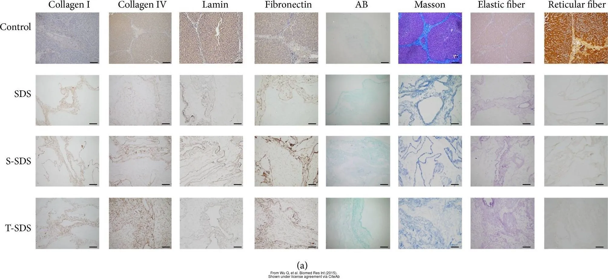
The data was published in the journal Biomed Res Int in 2015.
The data was published in the journal Biomed Res Int in 2015.
Fibronectin antibody
GTX72724
ApplicationsImmunoHistoChemistry, ImmunoHistoChemistry Paraffin
Product group Antibodies
ReactivityHuman, Porcine
TargetFN1
Overview
- SupplierGeneTex
- Product NameFibronectin antibody
- Delivery Days Customer9
- Application Supplier NoteImmunohistochemistry (IHC) 1:100-1:200 for 30 minute at room temperature using polymer system or streptavidin-biotin system. Formalin-fixed paraffin-embedded (FFPE) tissue sections and cell smears require antigen retriever, boiling tissue in 10 mM citrate buffer, pH 6.0 for 10-20 minute, followed by cooling at room temperature for 20 minutes.
- ApplicationsImmunoHistoChemistry, ImmunoHistoChemistry Paraffin
- CertificationResearch Use Only
- ClonalityPolyclonal
- Concentration2 mg/ml
- ConjugateUnconjugated
- Gene ID2335
- Target nameFN1
- Target descriptionfibronectin 1
- Target synonymsCIG, ED-B, FINC, FN, FNZ, GFND, GFND2, LETS, MSF, SMDCF, fibronectin, cold-insoluble globulin, epididymis secretory sperm binding protein, lnc-ABCA12-8, migration-stimulating factor
- HostRabbit
- IsotypeIgG
- Protein IDP02751
- Protein NameFibronectin
- Scientific DescriptionThis gene encodes fibronectin, a glycoprotein present in a soluble dimeric form in plasma, and in a dimeric or multimeric form at the cell surface and in extracellular matrix. The encoded preproprotein is proteolytically processed to generate the mature protein. Fibronectin is involved in cell adhesion and migration processes including embryogenesis, wound healing, blood coagulation, host defense, and metastasis. The gene has three regions subject to alternative splicing, with the potential to produce 20 different transcript variants, at least one of which encodes an isoform that undergoes proteolytic processing. The full-length nature of some variants has not been determined. [provided by RefSeq, Jan 2016]
- ReactivityHuman, Porcine
- Storage Instruction2°C to 8°C
- UNSPSC41116161
Datasheet
Related products
Product group Antibodies
Anti-FN1 AntibodyA36069
ApplicationsWestern Blot, ELISA, ImmunoHistoChemistry
ReactivityHuman, Mouse, Rat
- SizePrice
Product group Antibodies

Anti-Fibronectin [C6], Mouse IgG1, kappaAB04496-1.1
ApplicationsWestern Blot, ELISA, ImmunoHistoChemistry, Other Application
ReactivityHuman
TargetFN1
- SizePrice

Product group Antibodies
Anti-FN1 AntibodyAMAB91223
ApplicationsWestern Blot, ImmunoHistoChemistry
ReactivityHuman
TargetFN1
- SizePrice

Product group Antibodies
Anti-Fibronectin/FN1 Antibody Picoband(r)A00564-1-CARRIER-FREE
ApplicationsFlow Cytometry, Western Blot, ELISA, ImmunoHistoChemistry
ReactivityHuman
TargetFN1
- SizePrice
Product group Antibodies
Fibronectin Recombinant Antibody, AbBy Fluor-405 ConjugatedBSM-62010R-BF405
ApplicationsImmunoFluorescence, Western Blot
ReactivityHuman
TargetFN1
- SizePrice
Product group Antibodies
FN1 Monoclonal AntibodyCSB-MA000296
ApplicationsWestern Blot, ELISA
ReactivityHuman, Mouse, Rat
TargetFN1
- SizePrice
Product group Antibodies
ApplicationsImmunoPrecipitation, Western Blot, ImmunoCytoChemistry, ImmunoHistoChemistry
ReactivityPorcine
TargetFN1
- SizePrice
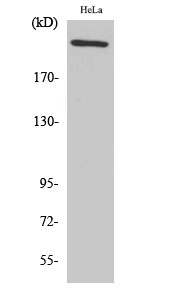
![WB analysis of 25 ug of human plasma lysates using GTX15737 Fibronectin antibody [3F12]. Dilution : 1:300](https://www.genetex.com/upload/website/prouct_img/normal/GTX15737/GTX15737_1519_WB_w_23060620_400.webp)
Product group Antibodies
Fibronectin antibody [3F12]GTX15737
ApplicationsImmunoFluorescence, ImmunoPrecipitation, Western Blot, ELISA, ImmunoCytoChemistry, RadioImmunoAssay
ReactivityHuman
TargetFN1
- SizePrice
Product group Antibodies
References
Fibronectin antibodyGTX20299
ApplicationsImmunoFluorescence, ImmunoPrecipitation, Western Blot, ELISA, ImmunoCytoChemistry, ImmunoHistoChemistry, ImmunoHistoChemistry Paraffin
ReactivityHuman
TargetFN1
- SizePrice