

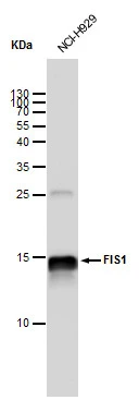
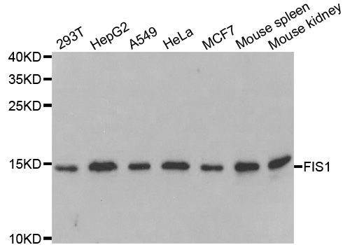
FIS1 antibody [GT12112] detects FIS1 protein by western blot analysis. Whole cell extracts (30 μg) was separated by 15 % SDS-PAGE, and blotted with FIS1 antibody [GT12112] (GTX630992) diluted by 1:1000
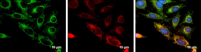
![FIS1 antibody [GT12112] detects FIS1 protein at mitochondria by immunofluorescent analysis. Sample: HeLa cells were fixed in 2% paraformaldehyde/culture medium at 37oC for 30 min. Green: FIS1 protein stained by FIS1 antibody [GT12112] (GTX630992) diluted at 1:500. Blue: Hoechst 33342 staining. Scale bar = 10 μm.](https://www.genetex.com/upload/website/prouct_img/normal/GTX630992/GTX630992_41750_IFA_w_23061202_854.webp "FIS1 antibody [GT12112] detects FIS1 protein at mitochondria by immunofluorescent analysis. Sample: HeLa cells were fixed in 2% paraformaldehyde/culture medium at 37oC for 30 min. Green: FIS1 protein stained by FIS1 antibody [GT12112] (GTX630992) diluted at 1:500. Blue: Hoechst 33342 staining. Scale bar = 10 μm.")
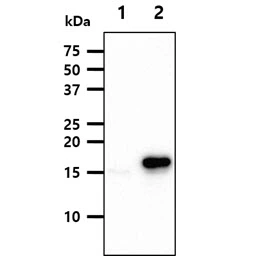
![Non-transfected (–) and transfected (+) HeLa whole cell extracts (30 μg) were separated by 15% SDS-PAGE, and the membrane was blotted with FIS1 antibody [GT12112] (GTX630992) diluted at 1:500.](https://www.genetex.com/upload/website/prouct_img/normal/GTX630992/GTX630992_41750_20161013_WB_shRNA_watermark_w_23061202_534.webp "Non-transfected (–) and transfected (+) HeLa whole cell extracts (30 μg) were separated by 15% SDS-PAGE, and the membrane was blotted with FIS1 antibody [GT12112] (GTX630992) diluted at 1:500.")
FIS1 antibody [GT12112] detects FIS1 protein by western blot analysis. Whole cell extracts (30 μg) was separated by 15 % SDS-PAGE, and blotted with FIS1 antibody [GT12112] (GTX630992) diluted by 1:1000
FIS1 antibody [GT12112]
GTX630992
ApplicationsImmunoFluorescence, Western Blot, ImmunoCytoChemistry
Product group Antibodies
ReactivityHuman
TargetFIS1
Overview
- SupplierGeneTex
- Product NameFIS1 antibody [GT12112]
- Delivery Days Customer9
- Application Supplier NoteWB: 1:500-1:3000. ICC/IF: 1:100-1:1000. *Optimal dilutions/concentrations should be determined by the researcher.Not tested in other applications.
- ApplicationsImmunoFluorescence, Western Blot, ImmunoCytoChemistry
- CertificationResearch Use Only
- ClonalityMonoclonal
- Clone IDGT12112
- Concentration3.55 mg/ml
- ConjugateUnconjugated
- Gene ID51024
- Target nameFIS1
- Target descriptionfission, mitochondrial 1
- Target synonymsCGI-135, TTC11, mitochondrial fission 1 protein, FIS1 homolog, H_NH0132A01.6, TPR repeat protein 11, fission 1 (mitochondrial outer membrane) homolog, hFis1, mitochondrial fission molecule, tetratricopeptide repeat domain 11, tetratricopeptide repeat protein 11
- HostMouse
- IsotypeIgG1
- Protein IDQ9Y3D6
- Protein NameMitochondrial fission 1 protein
- Scientific DescriptionThe balance between fission and fusion regulates the morphology of mitochondria. TTC11 is a component of a mitochondrial complex that promotes mitochondrial fission (James et al., 2003 [PubMed 12783892]).[supplied by OMIM]
- ReactivityHuman
- Storage Instruction-20°C or -80°C,2°C to 8°C
- UNSPSC12352203
Datasheet
Related products
Product group Antibodies
Anti-FIS1 (N-term) Antibody102-23904
ApplicationsWestern Blot
TargetFIS1
- SizePrice

Product group Antibodies
Anti-TTC11/FIS1 Antibody Picoband(r)A01932-2-CARRIER-FREE
ApplicationsFlow Cytometry, ImmunoFluorescence, Western Blot, ELISA, ImmunoCytoChemistry, ImmunoHistoChemistry
ReactivityHuman, Mouse, Rat
TargetFIS1
- SizePrice
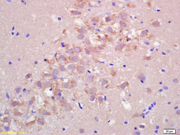
![IHC-P analysis of rat brain tissue section using GTX00950 FIS1 antibody [GT1188]. Dilution : 1:100](https://www.genetex.com/upload/website/prouct_img/normal/GTX00950/GTX00950_20200327_IHC-P_18_w_23053121_953.webp)
Product group Antibodies
FIS1 antibody [GT1188]GTX00950
ApplicationsImmunoFluorescence, ImmunoPrecipitation, Western Blot, ImmunoCytoChemistry, ImmunoHistoChemistry, ImmunoHistoChemistry Paraffin
ReactivityHuman, Mouse, Rat
TargetFIS1
- SizePrice
Product group Antibodies
References
FIS1 antibodyGTX111010
ApplicationsImmunoFluorescence, Western Blot, ImmunoCytoChemistry, ImmunoHistoChemistry, ImmunoHistoChemistry Paraffin
ReactivityHuman, Mouse, Rat
TargetFIS1
- SizePrice
Product group Antibodies
FIS1 antibody [AT3B7]GTX57722
ApplicationsImmunoFluorescence, Western Blot, ImmunoCytoChemistry
ReactivityHuman
TargetFIS1
- SizePrice
Product group Antibodies
FIS1 antibody [GT4211]GTX630983
ApplicationsImmunoFluorescence, Western Blot, ImmunoCytoChemistry
ReactivityHuman, Rat
TargetFIS1
- SizePrice
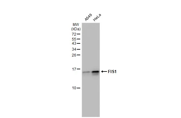
![Various whole cell extracts (30 μg) were separated by 15% SDS-PAGE, and the membrane was blotted with FIS1 antibody [GT9810] (GTX631209) diluted at 1:500. The HRP-conjugated anti-mouse IgG antibody (GTX213111-01) was used to detect the primary antibody.](https://www.genetex.com/upload/website/prouct_img/normal/GTX631209/GTX631209_41743_20191004_WB_R_w_23061202_369.webp)
Product group Antibodies
References
FIS1 antibody [GT9810]GTX631209
ApplicationsImmunoFluorescence, Western Blot, ImmunoCytoChemistry
ReactivityHuman, Mouse, Rat
TargetFIS1
- SizePrice
Product group Antibodies
Anti-FIS1 AntibodyA31072
ApplicationsImmunoFluorescence, Western Blot, ImmunoHistoChemistry
ReactivityHuman, Mouse, Rat
- SizePrice
Product group Antibodies
Fis1 Polyclonal AntibodyCAC07594
ApplicationsImmunoFluorescence, ELISA, ImmunoHistoChemistry
TargetFIS1
- SizePrice
Product group Antibodies
References
FIS1 Polyclonal AntibodyBS-7646R
ApplicationsImmunoFluorescence, Western Blot, ELISA, ImmunoCytoChemistry, ImmunoHistoChemistry, ImmunoHistoChemistry Frozen, ImmunoHistoChemistry Paraffin
ReactivityBovine, Canine, Equine, Human, Mouse, Porcine, Rat
TargetFIS1
- SizePrice