

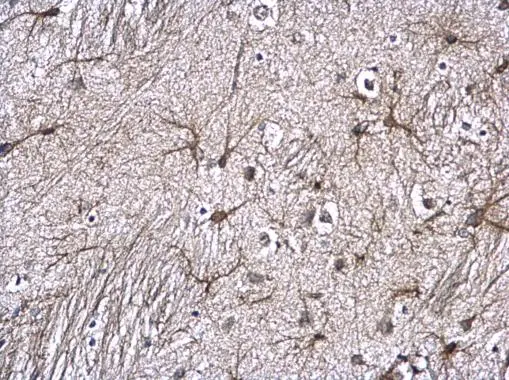
GFAP antibody detects GFAP protein at astrocyte on mouse fore brain by immunohistochemical analysis. Sample: Paraffin-embedded mouse fore brain. GFAP antibody (GTX100850) diluted at 1:500.
Antigen Retrieval: Trilogy? (EDTA based, pH 8.0) buffer, 15min
![GFAP antibody detects GFAP protein at glia cells by immunofluorescent analysis. Sample: DIV9 rat E18 primary cortical neurons were fixed in 4% paraformaldehyde at RT for 15 min. Green: GFAP protein stained by GFAP antibody (GTX100850) diluted at 1:500. Red: beta Tubulin 3/ Tuj1, a neuron cell marker, stained by beta Tubulin 3/ Tuj1 antibody [GT11710] (GTX631836) diluted at 1:500. Blue: Fluoroshield with DAPI (GTX30920).](https://www.genetex.com/upload/website/prouct_img/normal/GTX100850/GTX100850_42613_20170727_IFA_R_w_23060100_883.webp "GFAP antibody detects GFAP protein at glia cells by immunofluorescent analysis. Sample: DIV9 rat E18 primary cortical neurons were fixed in 4% paraformaldehyde at RT for 15 min. Green: GFAP protein stained by GFAP antibody (GTX100850) diluted at 1:500. Red: beta Tubulin 3/ Tuj1, a neuron cell marker, stained by beta Tubulin 3/ Tuj1 antibody [GT11710] (GTX631836) diluted at 1:500. Blue: Fluoroshield with DAPI (GTX30920).")
![GFAP antibody detects GFAP protein expression by immunohistochemical analysis. Sample: Frozen-sectioned adult mouse cerebellum. Green: GFAP protein stained by GFAP antibody (GTX100850) diluted at 1:250. Red: beta Tubulin 3/ TUJ1, stained by beta Tubulin 3/ TUJ1 antibody [GT11710] (GTX631836) diluted at 1:500. Blue: Fluoroshield with DAPI (GTX30920).](https://www.genetex.com/upload/website/prouct_img/normal/GTX100850/GTX100850_40268_20170531_IHC-Fr_M_w_23060100_158.webp "GFAP antibody detects GFAP protein expression by immunohistochemical analysis. Sample: Frozen-sectioned adult mouse cerebellum. Green: GFAP protein stained by GFAP antibody (GTX100850) diluted at 1:250. Red: beta Tubulin 3/ TUJ1, stained by beta Tubulin 3/ TUJ1 antibody [GT11710] (GTX631836) diluted at 1:500. Blue: Fluoroshield with DAPI (GTX30920).")
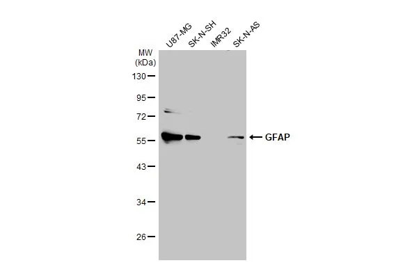
 and transfected (+) 293T whole cell extracts (30 μg) were separated by 10% SDS-PAGE, and the membrane was blotted with GFAP antibody (GTX100850) diluted at 1:2500. The HRP-conjugated anti-rabbit IgG antibody (GTX213110-01) was used to detect the primary antibody.")
![GFAP antibody detects GFAP protein at retinal ganglion cell layer by immunohistochemical analysis. Sample: Frozen sectioned adult mouse retina. Green: GFAP protein stained by GFAP antibody (GTX100850) diluted at 1:250. Red: beta Tubulin 3/ TUJ1, stained by beta Tubulin 3/ TUJ1 antibody [GT11710] (GTX631836) diluted at 1:250. Blue: Fluoroshield with DAPI (GTX30920).](https://www.genetex.com/upload/website/prouct_img/normal/GTX100850/GTX100850_40268_20160830_IHC-Fr_w_23060100_850.webp "GFAP antibody detects GFAP protein at retinal ganglion cell layer by immunohistochemical analysis. Sample: Frozen sectioned adult mouse retina. Green: GFAP protein stained by GFAP antibody (GTX100850) diluted at 1:250. Red: beta Tubulin 3/ TUJ1, stained by beta Tubulin 3/ TUJ1 antibody [GT11710] (GTX631836) diluted at 1:250. Blue: Fluoroshield with DAPI (GTX30920).")
. Green: GFAP antibody (GTX100850) diluted at 1:500. Blue: DAPI")
![GFAP antibodies detects GFAP proteins on embryonic mouse brain by immunohistochemical analysis. Sample: Frozen section of embryonic mouse brain (mE18.5). Green: GFAP antibody (GTX100850) diluted at 1:500. Red: Sox2 antibody [GT1876] (GTX627404) diluted at 1:500.](https://www.genetex.com/upload/website/prouct_img/normal/GTX100850/GTX100850_40268_20150625_IHC_M_2_w_23060100_866.webp "GFAP antibodies detects GFAP proteins on embryonic mouse brain by immunohistochemical analysis. Sample: Frozen section of embryonic mouse brain (mE18.5). Green: GFAP antibody (GTX100850) diluted at 1:500. Red: Sox2 antibody [GT1876] (GTX627404) diluted at 1:500.")
![GFAP antibody detects GFAP protein expression by immunohistochemical analysis. Sample: Frozen-sectioned adult mouse cerebellum. Green: GFAP protein stained by GFAP antibody (GTX100850) diluted at 1:250. Red: beta Tubulin 3/ TUJ1, stained by beta Tubulin 3/ TUJ1 antibody [GT11710] (GTX631836) diluted at 1:500. Blue: Fluoroshield with DAPI (GTX30920).](https://www.genetex.com/upload/website/prouct_img/normal/GTX100850/GTX100850_42613_20170531_IHC-Fr_M_w_23060100_657.webp "GFAP antibody detects GFAP protein expression by immunohistochemical analysis. Sample: Frozen-sectioned adult mouse cerebellum. Green: GFAP protein stained by GFAP antibody (GTX100850) diluted at 1:250. Red: beta Tubulin 3/ TUJ1, stained by beta Tubulin 3/ TUJ1 antibody [GT11710] (GTX631836) diluted at 1:500. Blue: Fluoroshield with DAPI (GTX30920).")
![GFAP antibody detects GFAP protein expression by immunohistochemical analysis. Sample: Frozen-sectioned adult mouse hippocampus. Green: GFAP protein stained by GFAP antibody (GTX100850) diluted at 1:250. Red: NeuN, stained by NeuN antibody [2Q158] (GTX30773) diluted at 1:500.](https://www.genetex.com/upload/website/prouct_img/normal/GTX100850/GTX100850_42613_42886_IHC-Fr_M_w_23060100_558.webp "GFAP antibody detects GFAP protein expression by immunohistochemical analysis. Sample: Frozen-sectioned adult mouse hippocampus. Green: GFAP protein stained by GFAP antibody (GTX100850) diluted at 1:250. Red: NeuN, stained by NeuN antibody [2Q158] (GTX30773) diluted at 1:500.")
 diluted at 1:1000. Antigen Retrieval: Citrate buffer, pH 6.0, 15 min")
GFAP antibody detects GFAP protein at astrocyte on mouse fore brain by immunohistochemical analysis. Sample: Paraffin-embedded mouse fore brain. GFAP antibody (GTX100850) diluted at 1:500.
Antigen Retrieval: Trilogy? (EDTA based, pH 8.0) buffer, 15min
GFAP antibody
GTX100850
ApplicationsImmunoFluorescence, Western Blot, ImmunoCytoChemistry, ImmunoHistoChemistry, ImmunoHistoChemistry Frozen, ImmunoHistoChemistry Paraffin
Product group Antibodies
ReactivityHuman, Mouse, Rat
TargetGFAP
Overview
- SupplierGeneTex
- Product NameGFAP antibody
- Delivery Days Customer9
- Application Supplier NoteWB: 1:500-1:3000. ICC/IF: 1:100-1:1000. IHC-P: 1:100-1:1000. IHC-Fr: 1:100-1:1000. *Optimal dilutions/concentrations should be determined by the researcher.Not tested in other applications.
- ApplicationsImmunoFluorescence, Western Blot, ImmunoCytoChemistry, ImmunoHistoChemistry, ImmunoHistoChemistry Frozen, ImmunoHistoChemistry Paraffin
- CertificationResearch Use Only
- ClonalityPolyclonal
- Concentration0.72 mg/ml
- ConjugateUnconjugated
- Gene ID2670
- Target nameGFAP
- Target descriptionglial fibrillary acidic protein
- Target synonymsALXDRD, glial fibrillary acidic protein
- HostRabbit
- IsotypeIgG
- Protein IDP14136
- Protein NameGlial fibrillary acidic protein
- Scientific DescriptionThis gene encodes one of the major intermediate filament proteins of mature astrocytes. It is used as a marker to distinguish astrocytes from other glial cells during development. Mutations in this gene cause Alexander disease, a rare disorder of astrocytes in the central nervous system. Alternative splicing results in multiple transcript variants encoding distinct isoforms. [provided by RefSeq]
- ReactivityHuman, Mouse, Rat
- Storage Instruction-20°C or -80°C,2°C to 8°C
- UNSPSC12352203
References
- Companys-Alemany J, Turcu AL, Vázquez S, et al. Glial cell reactivity and oxidative stress prevention in Alzheimer's disease mice model by an optimized NMDA receptor antagonist. Sci Rep. 2022,12(1):17908. doi: 10.1038/s41598-022-22963-xRead this paper
- Yang YM, Li CC, Yin le K, et al. Normalization of T2 relaxation time and apparent diffusion coefficient in relation to the inflammatory changes in the substantia nigra of rats with focal cerebral ischemia. Acta Radiol. 2015,56(7):837-43. doi: 10.1177/0284185114549496Read this paper
Datasheet
Related products
Product group Antibodies

Anti-GFAP R416WT [N206B/9]Ab02152-1.1
ApplicationsWestern Blot, ImmunoCytoChemistry, ImmunoHistoChemistry
ReactivityHuman, Mouse, Rat
TargetGFAP
- SizePrice
Product group Antibodies
Anti-GFAP Antibody130-10037
ApplicationsELISA
ReactivityHuman
TargetGFAP
- SizePrice

Product group Antibodies
Anti-GFAP AntibodyAMAB91033
ApplicationsWestern Blot, ImmunoHistoChemistry
ReactivityHuman, Mouse, Rat
TargetGFAP
- SizePrice
Product group Antibodies

ApplicationsImmunoFluorescence, Western Blot, ELISA, ImmunoCytoChemistry, ImmunoHistoChemistry
- SizePrice
Product group Antibodies
References
GFAP antibodyGTX108711
ApplicationsImmunoFluorescence, Western Blot, ImmunoCytoChemistry, ImmunoHistoChemistry, ImmunoHistoChemistry Frozen, ImmunoHistoChemistry Paraffin
ReactivityHuman, Mouse, Rat
TargetGFAP
- SizePrice
![GFAP antibody [HL1307] detects GFAP protein at cytoplasm by immunohistochemical analysis. Sample: Paraffin-embedded mouse brain. GFAP stained by GFAP antibody [HL1307] (GTX636725) diluted at 1:100. Antigen Retrieval: Citrate buffer, pH 6.0, 15 min](https://www.genetex.com/upload/website/prouct_img/normal/GTX636725/GTX636725_44627_20220408_IHC-P_M_22071401_482.webp)
Product group Antibodies
GFAP antibody [HL1307]GTX636725
ApplicationsWestern Blot, ImmunoHistoChemistry, ImmunoHistoChemistry Frozen, ImmunoHistoChemistry Paraffin
ReactivityHuman, Mouse, Rat
TargetGFAP
- SizePrice
![GFAP antibody [HL1308] detects GFAP protein at cytoplasm by immunohistochemical analysis. Sample: Paraffin-embedded rat brain. GFAP stained by GFAP antibody [HL1308] (GTX636726) diluted at 1:100. Antigen Retrieval: Citrate buffer, pH 6.0, 15 min](https://www.genetex.com/upload/website/prouct_img/normal/GTX636726/GTX636726_44627_20220408_IHC-P_R_22071401_888.webp)
Product group Antibodies
GFAP antibody [HL1308]GTX636726
ApplicationsImmunoFluorescence, ImmunoCytoChemistry, ImmunoHistoChemistry, ImmunoHistoChemistry Frozen, ImmunoHistoChemistry Paraffin
ReactivityHuman, Mouse, Rat
TargetGFAP
- SizePrice
Product group Antibodies
References
GFAP antibodyGTX85454
ApplicationsImmunoFluorescence, Western Blot, ImmunoCytoChemistry, ImmunoHistoChemistry, ImmunoHistoChemistry Frozen, ImmunoHistoChemistry Paraffin
ReactivityHuman, Mouse, Rat
TargetGFAP
- SizePrice
Product group Antibodies
Gfap Polyclonal AntibodyCAC08463
ApplicationsImmunoFluorescence, Western Blot, ELISA, ImmunoHistoChemistry
TargetGFAP
- SizePrice