

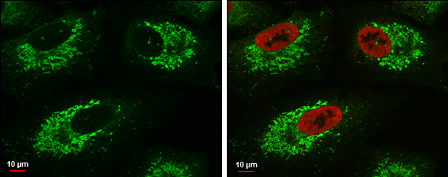
Glutaminase C antibody detects Glutaminase C protein at mitochondria by immunofluorescent analysis. Sample: A549 cells were fixed in 4% paraformaldehyde at RT for 15 min. Green: Glutaminase C protein stained by Glutaminase C antibody (GTX132402) diluted at 1:500. Red: Histone H3K9ac (acetyl Lys9), a nucleus marker, stained by Histone H3K9ac (acetyl Lys9) antibody [GT464] (GTX630554) diluted at 1:500. Blue: Hoechst 33342 staining. Scale bar = 10 μm.
 diluted at 1:500.")
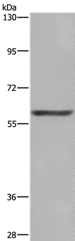
 antibody validation by shRNA knock-down. Non-transfected (-) and GLS1(GAC isoform specific) shRNA-transfected HCT116 whole cell extracts (30 μg) were separated by 10% SDS-PAGE, and the membrane was blotted with Glutaminase C (GAC) antibody (GTX132402) at a dilution of 1:5000.")
 or Glutaminase C antibody (GTX131263). Western blot analysis was performed using Glutaminase C antibody (GTX132402). EasyBlot anti-Rabbit IgG (GTX221666-01) was used as a secondary reagent.")
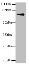
 and GLS1(GAC isoform specific) shRNA-transfected MDA-MB-231 whole cell extracts (30 μg) were separated by 7.5% SDS-PAGE, and the membrane was blotted with Glutaminase antibody (GTX132402) diluted by 1:5000.")
Glutaminase C antibody detects Glutaminase C protein at mitochondria by immunofluorescent analysis. Sample: A549 cells were fixed in 4% paraformaldehyde at RT for 15 min. Green: Glutaminase C protein stained by Glutaminase C antibody (GTX132402) diluted at 1:500. Red: Histone H3K9ac (acetyl Lys9), a nucleus marker, stained by Histone H3K9ac (acetyl Lys9) antibody [GT464] (GTX630554) diluted at 1:500. Blue: Hoechst 33342 staining. Scale bar = 10 μm.
Glutaminase C (GAC) antibody
GTX132402
ApplicationsImmunoFluorescence, ImmunoPrecipitation, Western Blot, ImmunoCytoChemistry, ImmunoHistoChemistry, ImmunoHistoChemistry Paraffin
Product group Antibodies
ReactivityHuman
TargetGLS
Overview
- SupplierGeneTex
- Product NameGlutaminase C (GAC) antibody
- Delivery Days Customer9
- Application Supplier NoteWB: 1:1000-1:10000. ICC/IF: 1:100-1:1000. IHC-P: 1:100-1:1000. IP: 1:100-1:500. *Optimal dilutions/concentrations should be determined by the researcher.Not tested in other applications.
- ApplicationsImmunoFluorescence, ImmunoPrecipitation, Western Blot, ImmunoCytoChemistry, ImmunoHistoChemistry, ImmunoHistoChemistry Paraffin
- CertificationResearch Use Only
- ClonalityPolyclonal
- Concentration0.96 mg/ml
- ConjugateUnconjugated
- Gene ID2744
- Target nameGLS
- Target descriptionglutaminase
- Target synonymsAAD20, CASGID, DEE71, EIEE71, GAC, GAM, GDPAG, GLS1, KGA, glutaminase kidney isoform, mitochondrial, K-glutaminase, L-glutamine amidohydrolase, glutaminase C, glutaminase, phosphate-activated
- HostRabbit
- IsotypeIgG
- Protein IDO94925
- Protein NameGlutaminase kidney isoform, mitochondrial
- Scientific DescriptionSahai (1983) [PubMed 6825316] demonstrated phosphate-activated glutaminase (EC 3.5.1.2) in human platelets. It is the major enzyme yielding glutamate from glutamine. Significance of the enzyme derives from its possible implication in behavior disturbances in which glutamate acts as a neurotransmitter (Prusiner, 1981). High heritability of platelet glutaminase was indicated by studies of Sahai and Vogel (1983) [PubMed 6682827] who found an intraclass correlation coefficient of 0.96 for monozygotic twins and 0.53 for dizygotic twins.[supplied by OMIM]
- ReactivityHuman
- Storage Instruction-20°C or -80°C,2°C to 8°C
- UNSPSC41116161
Datasheet
Related products
Product group Antibodies
Anti-GLS AntibodyA39155
ApplicationsWestern Blot, ImmunoHistoChemistry
ReactivityHuman
- SizePrice

Product group Antibodies
Anti-Glutaminase/GLS Antibody Picoband(r)A01272-2-CARRIER-FREE
ApplicationsFlow Cytometry, ImmunoFluorescence, Western Blot, ELISA, ImmunoCytoChemistry, ImmunoHistoChemistry
ReactivityHuman, Monkey, Mouse, Rat
TargetGLS
- SizePrice
Product group Antibodies
Anti-GLS Antibody144-03885
ApplicationsWestern Blot, ImmunoHistoChemistry
ReactivityHuman, Mouse, Rat
TargetGLS
- SizePrice

Product group Antibodies

GLS / Glutaminase AntibodyLS-C831065
ApplicationsWestern Blot, ELISA, ImmunoHistoChemistry
ReactivityHuman, Mouse, Rat
TargetGLS
- SizePrice
Product group Antibodies
GLS1 Polyclonal AntibodyBS-10341R
ApplicationsImmunoFluorescence, Western Blot, ELISA, ImmunoHistoChemistry, ImmunoHistoChemistry Frozen, ImmunoHistoChemistry Paraffin
ReactivityHuman, Mouse, Rat
TargetGLS
- SizePrice
Product group Antibodies
GLS AntibodyCSB-PA009528DA01HU
ApplicationsWestern Blot, ELISA, ImmunoHistoChemistry
ReactivityHuman, Mouse
TargetGLS
- SizePrice
Product group Antibodies
GLS Polyclonal AntibodyCAC13047
ApplicationsWestern Blot, ELISA, ImmunoHistoChemistry
ReactivityMouse
TargetGLS
- SizePrice
Product group Antibodies
References
Glutaminase antibody, C-termGTX81012
ApplicationsFlow Cytometry, ImmunoFluorescence, Western Blot, ImmunoCytoChemistry, ImmunoHistoChemistry, ImmunoHistoChemistry Paraffin
ReactivityHuman, Mouse, Rat
TargetGLS
- SizePrice