

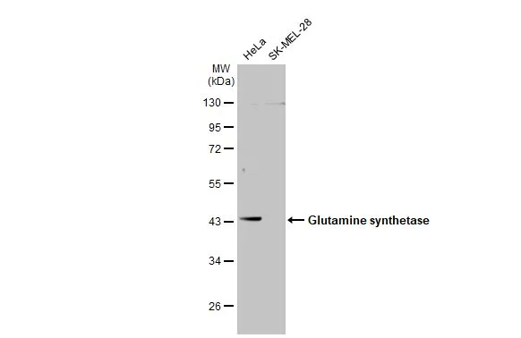
Various whole cell extracts (30 μg) were separated by 10% SDS-PAGE, and the membrane was blotted with Glutamine synthetase antibody (GTX109121) diluted at 1:1000. The HRP-conjugated anti-rabbit IgG antibody (GTX213110-01) was used to detect the primary antibody.
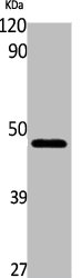
 was separated by 10% SDS-PAGE, and the membrane was blotted with Glutamine synthetase antibody (GTX109121) diluted at 1:10000.")
 dilution: 1:100.")
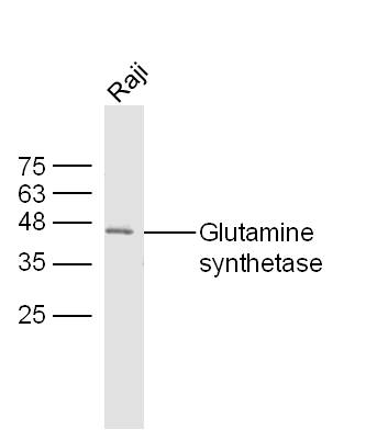
 was separated by 10 % SDS-PAGE, and the membrane was blotted with Glutamine synthetase antibody (GTX109121) at a dilution of 1:10000.")
 dilution: 1:100.")
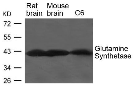
 was separated by 10% SDS-PAGE, and the membrane was blotted with Glutamine synthetase antibody (GTX109121) diluted at 1:50000.")
 was separated by 10% SDS-PAGE, and the membrane was blotted with Glutamine synthetase antibody (GTX109121) diluted at 1:20000.")
 was separated by 10% SDS-PAGE, and the membrane was blotted with Glutamine synthetase antibody (GTX109121) diluted at 1:50000.")
![Glutamine synthetase antibody detects Glutamine synthetase protein expression by immunohistochemical analysis. Sample:Paraffin-embedded adult mouse retina. Green: Glutamine synthetase protein stained by Glutamine synthetase antibody (GTX109121) diluted at 1:250. Red: beta Tubulin 3/ TUJ1, stained by beta Tubulin 3/ TUJ1 antibody [GT11710] (GTX631836) diluted at 1:250. Blue: Fluoroshield with DAPI (GTX30920).](https://www.genetex.com/upload/website/prouct_img/normal/GTX109121/GTX109121_41745_20170214_IHC-P_w_23060120_113.webp "Glutamine synthetase antibody detects Glutamine synthetase protein expression by immunohistochemical analysis. Sample:Paraffin-embedded adult mouse retina. Green: Glutamine synthetase protein stained by Glutamine synthetase antibody (GTX109121) diluted at 1:250. Red: beta Tubulin 3/ TUJ1, stained by beta Tubulin 3/ TUJ1 antibody [GT11710] (GTX631836) diluted at 1:250. Blue: Fluoroshield with DAPI (GTX30920).")
![Glutamine synthetase antibody detects Glutamine synthetase protein expression by immunohistochemical analysis. Sample: Frozen-sectioned adult mouse cerebellum. Green: Glutamine synthetase protein stained by Glutamine synthetase antibody (GTX109121) diluted at 1:250. Red: beta Tubulin 3/ TUJ1, stained by beta Tubulin 3/ TUJ1 antibody [GT11710] (GTX631836) diluted at 1:500. Blue: Fluoroshield with DAPI (GTX30920).](https://www.genetex.com/upload/website/prouct_img/normal/GTX109121/GTX109121_41626_20170531_IHC-Fr_M_w_23060120_821.webp "Glutamine synthetase antibody detects Glutamine synthetase protein expression by immunohistochemical analysis. Sample: Frozen-sectioned adult mouse cerebellum. Green: Glutamine synthetase protein stained by Glutamine synthetase antibody (GTX109121) diluted at 1:250. Red: beta Tubulin 3/ TUJ1, stained by beta Tubulin 3/ TUJ1 antibody [GT11710] (GTX631836) diluted at 1:500. Blue: Fluoroshield with DAPI (GTX30920).")
Various whole cell extracts (30 μg) were separated by 10% SDS-PAGE, and the membrane was blotted with Glutamine synthetase antibody (GTX109121) diluted at 1:1000. The HRP-conjugated anti-rabbit IgG antibody (GTX213110-01) was used to detect the primary antibody.
Glutamine synthetase antibody
GTX109121
ApplicationsImmunoFluorescence, ImmunoPrecipitation, Western Blot, ImmunoCytoChemistry, ImmunoHistoChemistry, ImmunoHistoChemistry Frozen, ImmunoHistoChemistry Paraffin
Product group Antibodies
ReactivityHuman, Mouse, Rat, Zebra Fish
TargetGLUL
Overview
- SupplierGeneTex
- Product NameGlutamine synthetase antibody
- Delivery Days Customer9
- Application Supplier NoteWB: 1:5000-1:20000. ICC/IF: 1:100-1:1000. IHC-P: 1:100-1:1000. IHC-Fr: 1:100-1:1000. IP: 1:100-1:500. *Optimal dilutions/concentrations should be determined by the researcher.Not tested in other applications.
- ApplicationsImmunoFluorescence, ImmunoPrecipitation, Western Blot, ImmunoCytoChemistry, ImmunoHistoChemistry, ImmunoHistoChemistry Frozen, ImmunoHistoChemistry Paraffin
- CertificationResearch Use Only
- ClonalityPolyclonal
- Concentration0.72 mg/ml
- ConjugateUnconjugated
- Gene ID2752
- Target nameGLUL
- Target descriptionglutamate-ammonia ligase
- Target synonymsDEE116, GLNS, GS, PIG43, PIG59, glutamine synthetase, cell proliferation-inducing protein 59, glutamate decarboxylase, glutamine synthase, palmitoyltransferase GLUL, proliferation-inducing protein 43
- HostRabbit
- IsotypeIgG
- Protein IDP15104
- Protein NameGlutamine synthetase
- Scientific DescriptionGlutamine is a main source of energy and is involved in cell proliferation, inhibition of apoptosis, and cell signaling (Haberle et al., 2005 [PubMed 16267323]). Fetal glutamine requirements are very high and depend largely on active glutamine synthesis and the release of glutamine into the fetal circulation by the placenta. Glutamine synthetase (EC 6.3.1.2), also called glutamate-ammonia ligase (GLUL), is expressed throughout the body and plays an important role in controlling body pH and in removing ammonia from the circulation. The enzyme clears L-glutamate, the major neurotransmitter in the central nervous system, from neuronal synapses (see references in Clancy et al., 1996 [PubMed 8975719]).[supplied by OMIM]
- ReactivityHuman, Mouse, Rat, Zebra Fish
- Storage Instruction-20°C or -80°C,2°C to 8°C
- UNSPSC41116161
Datasheet
Related products

Product group Antibodies
Anti-GLUL Antibody Picoband(r)A03191-3-CARRIER-FREE
ApplicationsFlow Cytometry, ImmunoFluorescence, Western Blot, ELISA, ImmunoCytoChemistry, ImmunoHistoChemistry
ReactivityHuman, Mouse, Rat
TargetGLUL
- SizePrice
Product group Antibodies
Anti-GLUL Antibody144-05437
ApplicationsImmunoFluorescence, Western Blot, ImmunoHistoChemistry
ReactivityHuman, Mouse
TargetGLUL
- SizePrice

Product group Antibodies
Anti-GLUL AntibodyAMAB91101
ApplicationsWestern Blot, ImmunoHistoChemistry
ReactivityHuman, Mouse, Rat
TargetGLUL
- SizePrice
Product group Antibodies
ApplicationsWestern Blot
ReactivityHuman, Mouse, Rat
- SizePrice
Product group Antibodies
ApplicationsFlow Cytometry, ImmunoFluorescence, Western Blot, ELISA, ImmunoCytoChemistry, ImmunoHistoChemistry, ImmunoHistoChemistry Frozen, ImmunoHistoChemistry Paraffin
ReactivityBovine, Human, Mouse, Porcine, Rat, Sheep
TargetGLUL
- SizePrice
Product group Antibodies
GLUL Polyclonal AntibodyCAC14536
ApplicationsImmunoFluorescence, Western Blot, ELISA, ImmunoHistoChemistry
ReactivityMouse
TargetGLUL
- SizePrice
Product group Antibodies
GLUL AntibodyCSB-PA004642
ApplicationsWestern Blot, ELISA
ReactivityHuman, Mouse, Rat
TargetGLUL
- SizePrice
Product group Antibodies

GLUL / Glutamine Synthetase AntibodyLS-C401718
ApplicationsWestern Blot, ELISA, ImmunoHistoChemistry
ReactivityHuman, Mouse, Rat
TargetGLUL
- SizePrice
![IHC-P analysis of human liver tissue using GTX04471 Glutamine synthetase antibody [MSVA-750M] HistoMAX?. Glutamine synthetase staining is strong in centrilobular hepatocytes weak to moderate in Kupffer cells but absent in periportal hepatocytes.](https://www.genetex.com/upload/website/prouct_img/normal/GTX04471/GTX04471_20230728_IHC-P_57_23072722_662.webp)
Product group Antibodies
ApplicationsImmunoHistoChemistry, ImmunoHistoChemistry Paraffin
ReactivityHuman
TargetGLUL
- SizePrice
![IHC-P analysis of human endometrium tissue using GTX84426 Glutamine synthetase antibody [1F4]. Antigen retrieval : Heat-induced epitope retrieval by 10mM citrate buffer, pH6.0, 100oC for 10min.](https://www.genetex.com/upload/website/prouct_img/normal/GTX84426/GTX84426_2705_IHC-P_w_23061420_245.webp)
Product group Antibodies
References
ApplicationsFlow Cytometry, ImmunoFluorescence, Western Blot, ImmunoCytoChemistry, ImmunoHistoChemistry, ImmunoHistoChemistry Paraffin
ReactivityHuman
TargetGLUL
- SizePrice