

Goat anti-SATB1
EB09448
ApplicationsFlow Cytometry, ImmunoFluorescence, Western Blot, ELISA, ImmunoHistoChemistry
Product group Antibodies
ReactivityBovine, Human, Mouse, Rat
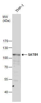
TargetSATB1
Overview
- SupplierEverest Biotech
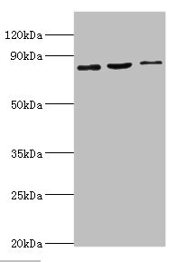
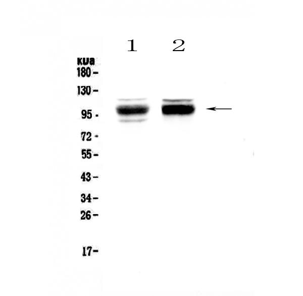
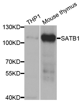
- Product NameGoat anti-SATB1
- Delivery Days Customer5
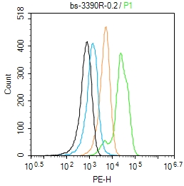
- Application Supplier NoteImmunofluorescence: Strong expression of the protein seen in the nuclei of Jurkat cells. Recommended concentration: 10microg/ml. Flow Cytometry: Flow cytometric analysis of Jurkat cells. Recommended concentration: 10ug/ml.
- ApplicationsFlow Cytometry, ImmunoFluorescence, Western Blot, ELISA, ImmunoHistoChemistry
- Applications SupplierPep-ELISA, WB, IHC, IF, FC
- CertificationResearch Use Only
- ClonalityPolyclonal
- Concentration0.5 mg/ml
- Gene ID6304
- Target nameSATB1
- Target descriptionSATB homeobox 1
- Target synonymsDEFDA, DHDBV, KTZSL, DNA-binding protein SATB1, special AT-rich sequence binding protein 1 (binds to nuclear matrix/scaffold-associating DNA)
- HostGoat
- Scientific DescriptionRefSeq number(s): NP_002962.1; NP_001182399.1; NP_001309805.1. GeneIDs all Nonhuman: 20230 (mouse); 316164 (rat);. Purification: Antigen affinity purified. Names and symbols: SATB1; SATB homeobox 1; DNA-binding protein SATB1; special AT-rich sequence binding protein 1; special AT-rich sequence binding protein 1 (binds to nuclear matrix/scaffold-associating DNA's)
- ReactivityBovine, Human, Mouse, Rat
- Reactivity SupplierHuman, Mouse, Rat, Cow
- Storage Instruction-20°C
- UNSPSC12352203
Related products
Product group Antibodies
SATB1 AntibodyCSB-PA020719DSR1HU
ApplicationsWestern Blot, ELISA
ReactivityHuman, Mouse
TargetSATB1
- SizePrice
Product group Antibodies
Anti-SATB1 Antibody Picoband(r)A01312-1-CARRIER-FREE
ApplicationsFlow Cytometry, Western Blot, ELISA, ImmunoCytoChemistry, ImmunoHistoChemistry
ReactivityHuman, Mouse, Rat
TargetSATB1
- SizePrice
Product group Antibodies
Anti-SATB1 AntibodyA31058
ApplicationsImmunoFluorescence, Western Blot, ImmunoHistoChemistry
ReactivityHuman, Mouse, Rat
- SizePrice

Product group Antibodies
Anti-SATB1 AntibodyHPA051769
ApplicationsImmunoCytoChemistry, ImmunoHistoChemistry
ReactivityHuman
TargetSATB1
- SizePrice
Product group Antibodies

SATB1 AntibodyLS-C334301
ApplicationsImmunoFluorescence, Western Blot, ImmunoHistoChemistry
ReactivityHuman, Mouse, Rat
TargetSATB1
- SizePrice
Product group Antibodies
ApplicationsImmunoPrecipitation, Western Blot, ImmunoCytoChemistry, ImmunoHistoChemistry
TargetSATB1
- SizePrice
Product group Antibodies
ApplicationsFlow Cytometry, ImmunoFluorescence, ELISA, ImmunoCytoChemistry, ImmunoHistoChemistry, ImmunoHistoChemistry Frozen, ImmunoHistoChemistry Paraffin
ReactivityBovine, Canine, Chicken, Human, Mouse, Porcine, Rabbit
TargetSATB1
- SizePrice
Product group Antibodies
SATB1 antibodyGTX114737
ApplicationsWestern Blot, ImmunoHistoChemistry, ImmunoHistoChemistry Paraffin
ReactivityHuman, Mouse, Rat
TargetSATB1
- SizePrice
Product group Antibodies
Anti-SATB1 Antibody144-05800
ApplicationsWestern Blot, ImmunoHistoChemistry
ReactivityHuman, Mouse, Rat
TargetSATB1
- SizePrice