

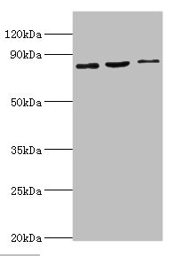
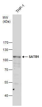
Western blot All lanes: SATB1 antibody at 2ug/ml Lane 1: Mouse brain tissue Lane 2: 293T whole cell lysate Lane 3: Jurkat whole cell lysate Secondary Goat polyclonal to rabbit IgG at 1/10000 dilution Predicted band size: 86, 90 kDa Observed band size: 86 kDa
Western blot All lanes: SATB1 antibody at 2ug/ml Lane 1: Mouse brain tissue Lane 2: 293T whole cell lysate Lane 3: Jurkat whole cell lysate Secondary Goat polyclonal to rabbit IgG at 1/10000 dilution Predicted band size: 86, 90 kDa Observed band size: 86 kDa
SATB1 Antibody
CSB-PA020719DSR1HU
ApplicationsWestern Blot, ELISA
Product group Antibodies
ReactivityHuman, Mouse
TargetSATB1
Overview
- SupplierCusabio
- Product NameSATB1 Antibody
- Delivery Days Customer20
- ApplicationsWestern Blot, ELISA
- CertificationResearch Use Only
- ClonalityPolyclonal
- ConjugateUnconjugated
- Gene ID6304
- Target nameSATB1
- Target descriptionSATB homeobox 1
- Target synonymsDEFDA, DHDBV, KTZSL, DNA-binding protein SATB1, special AT-rich sequence binding protein 1 (binds to nuclear matrix/scaffold-associating DNA)
- HostRabbit
- IsotypeIgG
- Protein IDQ01826
- Protein NameDNA-binding protein SATB1
- Scientific DescriptionCrucial silencing factor contributing to the initiation of X inactivation mediated by Xist RNA that occurs during embryogenesis and in lymphoma (By similarity). Binds to DNA at special AT-rich sequences, the consensus SATB1-binding sequence (CSBS), at nuclear matrix- or scaffold-associated regions. Thought to recognize the sugar-phosphate structure of double-stranded DNA. Transcriptional repressor controlling nuclear and viral gene expression in a phosphorylated and acetylated status-dependent manner, by binding to matrix attachment regions (MARs) of DNA and inducing a local chromatin-loop remodeling. Acts as a docking site for several chromatin remodeling enzymes (e.g. PML at the MHC-I locus) and also by recruiting corepressors (HDACs) or coactivators (HATs) directly to promoters and enhancers. Modulates genes that are essential in the maturation of the immune T-cell CD8SP from thymocytes. Required for the switching of fetal globin species, and beta- and gamma-globin genes regulation during erythroid differentiation. Plays a role in chromatin organization and nuclear architecture during apoptosis. Interacts with the unique region (UR) of cytomegalovirus (CMV). Alu-like motifs and SATB1-binding sites provide a unique chromatin context which seems preferentially targeted by the HIV-1 integration machinery. Moreover, HIV-1 Tat may overcome SATB1-mediated repression of IL2 and IL2RA (interleukin) in T-cells by binding to the same domain than HDAC1. Delineates specific epigenetic modifications at target gene loci, directly up-regulating metastasis-associated genes while down-regulating tumor-suppressor genes. Reprograms chromatin organization and the transcription profiles of breast tumors to promote growth and metastasis.
- ReactivityHuman, Mouse
- Storage Instruction-20°C or -80°C
- UNSPSC41116161
Related products
Product group Antibodies
Anti-SATB1 AntibodyA31058
ApplicationsImmunoFluorescence, Western Blot, ImmunoHistoChemistry
ReactivityHuman, Mouse, Rat
- SizePrice
Product group Antibodies
Anti-SATB1 Antibody Picoband(r)A01312-1-CARRIER-FREE
ApplicationsFlow Cytometry, Western Blot, ELISA, ImmunoCytoChemistry, ImmunoHistoChemistry
ReactivityHuman, Mouse, Rat
TargetSATB1
- SizePrice
Product group Antibodies
Anti-SATB1 Antibody144-05800
ApplicationsWestern Blot, ImmunoHistoChemistry
ReactivityHuman, Mouse, Rat
TargetSATB1
- SizePrice
Product group Antibodies
ApplicationsFlow Cytometry, ImmunoFluorescence, ELISA, ImmunoCytoChemistry, ImmunoHistoChemistry, ImmunoHistoChemistry Frozen, ImmunoHistoChemistry Paraffin
ReactivityBovine, Canine, Chicken, Human, Mouse, Porcine, Rabbit
TargetSATB1
- SizePrice
Product group Antibodies

Goat anti-SATB1EB09448
ApplicationsFlow Cytometry, ImmunoFluorescence, Western Blot, ELISA, ImmunoHistoChemistry
ReactivityBovine, Human, Mouse, Rat
TargetSATB1
- SizePrice
Product group Antibodies
ApplicationsImmunoPrecipitation, Western Blot, ImmunoCytoChemistry, ImmunoHistoChemistry
TargetSATB1
- SizePrice
Product group Antibodies

SATB1 AntibodyLS-C334301
ApplicationsImmunoFluorescence, Western Blot, ImmunoHistoChemistry
ReactivityHuman, Mouse, Rat
TargetSATB1
- SizePrice

Product group Antibodies
Anti-SATB1 AntibodyHPA051769
ApplicationsImmunoCytoChemistry, ImmunoHistoChemistry
ReactivityHuman
TargetSATB1
- SizePrice
Product group Antibodies
SATB1 antibodyGTX114737
ApplicationsWestern Blot, ImmunoHistoChemistry, ImmunoHistoChemistry Paraffin
ReactivityHuman, Mouse, Rat
TargetSATB1
- SizePrice