

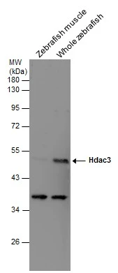
Various tissue extracts (30 μg) were separated by 10% SDS-PAGE, and the membrane was blotted with HDAC3 antibody (GTX113303) diluted at 1:500.
 at 1:300 dilution.")
 of zebrafish embryo, using HDAC3 antibody (GTX113303) at 1:200 dilution.")
 were separated by 10% SDS-PAGE, and the membrane was blotted with HDAC3 antibody (GTX113303) diluted by 1:1000.")
![HDAC3 antibody detects HDAC3 protein at nucleus by immunofluorescent analysis. Sample: HeLa cells were fixed in 4% paraformaldehyde at RT for 15 min. Green: HDAC3 protein stained by HDAC3 antibody (GTX113303) diluted at 1:1000. Red: alpha Tubulin, a cytoskeleton marker, stained by alpha Tubulin antibody [GT114] (GTX628802) diluted at 1:1000. Blue: Hoechst 33342 staining.](https://www.genetex.com/upload/website/prouct_img/normal/GTX113303/GTX113303_40436_20150410_IFA_w_23060501_438.webp "HDAC3 antibody detects HDAC3 protein at nucleus by immunofluorescent analysis. Sample: HeLa cells were fixed in 4% paraformaldehyde at RT for 15 min. Green: HDAC3 protein stained by HDAC3 antibody (GTX113303) diluted at 1:1000. Red: alpha Tubulin, a cytoskeleton marker, stained by alpha Tubulin antibody [GT114] (GTX628802) diluted at 1:1000. Blue: Hoechst 33342 staining.")
 were separated by 10% SDS-PAGE, and the membrane was blotted with HDAC3 antibody (GTX113303) diluted at 1:500. The HRP-conjugated anti-rabbit IgG antibody (GTX213110-01) was used to detect the primary antibody.")
 were separated by 10% SDS-PAGE, and the membrane was blotted with HDAC3 antibody (GTX113303) diluted at 1:1000. The HRP-conjugated anti-rabbit IgG antibody (GTX213110-01) was used to detect the primary antibody.")

 7.5% SDS-PAGE The immunoprecipitated HDAC3 protein was detected by HDAC3 antibody (GTX113303) diluted at 1:1000. EasyBlot anti-rabbit IgG (GTX221666-01) was used as a secondary reagent.")
 were separated by 10% SDS-PAGE, and the membrane was blotted with HDAC3 antibody (GTX113303) diluted at 1:1000. The HRP-conjugated anti-rabbit IgG antibody (GTX213110-01) was used to detect the primary antibody.")
Various tissue extracts (30 μg) were separated by 10% SDS-PAGE, and the membrane was blotted with HDAC3 antibody (GTX113303) diluted at 1:500.
HDAC3 antibody
GTX113303
ApplicationsImmunoFluorescence, ImmunoPrecipitation, Western Blot, ChIP Chromatin ImmunoPrecipitation, ImmunoCytoChemistry, ImmunoHistoChemistry, ImmunoHistoChemistry Paraffin
Product group Antibodies
ReactivityHuman, Mouse, Rat, Zebra Fish
TargetHDAC3
Overview
- SupplierGeneTex
- Product NameHDAC3 antibody
- Delivery Days Customer9
- Application Supplier NoteWB: 1:500-1:3000. ICC/IF: 1:100-1:1000. IHC-P: 1:100-1:1000. IP: 1:100-1:500. *Optimal dilutions/concentrations should be determined by the researcher.Not tested in other applications.
- ApplicationsImmunoFluorescence, ImmunoPrecipitation, Western Blot, ChIP Chromatin ImmunoPrecipitation, ImmunoCytoChemistry, ImmunoHistoChemistry, ImmunoHistoChemistry Paraffin
- CertificationResearch Use Only
- ClonalityPolyclonal
- Concentration0.32 mg/ml
- ConjugateUnconjugated
- Gene ID8841
- Target nameHDAC3
- Target descriptionhistone deacetylase 3
- Target synonymsHD3, KDAC3, RPD3, RPD3-2, histone deacetylase 3, SMAP45, protein deacetylase HDAC3, protein deacylase HDAC3
- HostRabbit
- IsotypeIgG
- Protein IDO15379
- Protein NameHistone deacetylase 3
- Scientific DescriptionHistones play a critical role in transcriptional regulation, cell cycle progression, and developmental events. Histone acetylation/deacetylation alters chromosome structure and affects transcription factor access to DNA. The protein encoded by this gene belongs to the histone deacetylase/acuc/apha family. It has histone deacetylase activity and represses transcription when tethered to a promoter. It may participate in the regulation of transcription through its binding with the zinc-finger transcription factor YY1. This protein can also down-regulate p53 function and thus modulate cell growth and apoptosis. This gene is regarded as a potential tumor suppressor gene. [provided by RefSeq]
- ReactivityHuman, Mouse, Rat, Zebra Fish
- Storage Instruction-20°C or -80°C,2°C to 8°C
- UNSPSC12352203
References
- Martirosian V, Deshpande K, Zhou H, et al. Medulloblastoma uses GABA transaminase to survive in the cerebrospinal fluid microenvironment and promote leptomeningeal dissemination. Cell Rep. 2021,35(13):109302. doi: 10.1016/j.celrep.2021.109302Read this paper
- Miscianinov V, Martello A, Rose L, et al. MicroRNA-148b Targets the TGF-β Pathway to Regulate Angiogenesis and Endothelial-to-Mesenchymal Transition during Skin Wound Healing. Mol Ther. 2018,26(8):1996-2007. doi: 10.1016/j.ymthe.2018.05.002Read this paper
- Armour SM, Remsberg JR, Damle M, et al. An HDAC3-PROX1 corepressor module acts on HNF4α to control hepatic triglycerides. Nat Commun. 2017,8(1):549. doi: 10.1038/s41467-017-00772-5Read this paper
- Emmett MJ, Lim HW, Jager J, et al. Histone deacetylase 3 prepares brown adipose tissue for acute thermogenic challenge. Nature. 2017,546(7659):544-548. doi: 10.1038/nature22819Read this paper
- Soriano FX, Hardingham GE. In cortical neurons HDAC3 activity suppresses RD4-dependent SMRT export. PLoS One. 2011,6(6):e21056. doi: 10.1371/journal.pone.0021056Read this paper
Datasheet
Related products
Product group Antibodies
ApplicationsImmunoFluorescence, ChIP Chromatin ImmunoPrecipitation
ReactivityHuman
TargetHDAC3
- SizePrice
Product group Antibodies
Anti-HDAC3 Antibody144-02139
ApplicationsImmunoFluorescence, ImmunoPrecipitation, Western Blot, ChIP Chromatin ImmunoPrecipitation, ImmunoHistoChemistry
ReactivityHuman, Mouse, Rat
TargetHDAC3
- SizePrice

Product group Antibodies
Anti-HDAC3 Antibody Picoband(r)A00839-CARRIER-FREE
ApplicationsFlow Cytometry, Western Blot, ELISA
ReactivityHuman, Mouse, Rat
TargetHDAC3
- SizePrice
![Various whole cell extracts (30 μg) were separated by 10% SDS-PAGE, and the membrane was blotted with HDAC3 antibody [GT1144] (GTX00834) diluted at 1:1000. The HRP-conjugated anti-rabbit IgG antibody (GTX213110-01) was used to detect the primary antibody, and the signal was developed with Trident ECL plus-Enhanced.](https://www.genetex.com/upload/website/prouct_img/normal/GTX00834/GTX00834_4000000016_20200410_WB_w_23053121_498.webp)
Product group Antibodies
HDAC3 antibody [GT1144]GTX00834
ApplicationsWestern Blot
ReactivityHuman, Mouse, Rat
TargetHDAC3
- SizePrice
![Whole cell extract (30 μg) was separated by 10% SDS-PAGE, and the membrane was blotted with HDAC3 antibody [C3], C-term (GTX109679) diluted at 1:500. The HRP-conjugated anti-rabbit IgG antibody (GTX213110-01) was used to detect the primary antibody.](https://www.genetex.com/upload/website/prouct_img/normal/GTX109679/GTX109679_44587_20220520_WB_23032819_175.webp)
Product group Antibodies
References
HDAC3 antibody [C3], C-termGTX109679
ApplicationsImmunoFluorescence, ImmunoPrecipitation, Western Blot, ChIP Chromatin ImmunoPrecipitation, ImmunoCytoChemistry, ImmunoHistoChemistry, ImmunoHistoChemistry Paraffin
ReactivityDrosophila, Human, Mouse, Rat
TargetHDAC3
- SizePrice
![ICC/IF analysis of 4% paraformaldehyde fixed HeLa cells using GTX60366 HDAC3 antibody [GT9007]. Red : Primary antibody Blue : DAPI Dilution : 1:500](https://www.genetex.com/upload/website/prouct_img/normal/GTX60366/GTX60366_20201117_ICCIF_153_w_23061123_371.webp)
Product group Antibodies
HDAC3 antibody [GT9007]GTX60366
ApplicationsImmunoFluorescence, ChIP Chromatin ImmunoPrecipitation, ImmunoCytoChemistry
ReactivityHuman
TargetHDAC3
- SizePrice
![Various whole cell extracts (30 μg) were separated by 10% SDS-PAGE, and the membrane was blotted with HDAC3 antibody [HL1749] (GTX637399) diluted at 1:1000. The HRP-conjugated anti-rabbit IgG antibody (GTX213110-01) was used to detect the primary antibody.](https://www.genetex.com/upload/website/prouct_img/normal/GTX637399/GTX637399_44837_20221021_WB_22102723_152.webp)
Product group Antibodies
HDAC3 antibody [HL1749]GTX637399
ApplicationsWestern Blot, ImmunoHistoChemistry, ImmunoHistoChemistry Paraffin
ReactivityDrosophila, Human, Mouse, Rat
TargetHDAC3
- SizePrice
Product group Antibodies
HDAC3 Polyclonal AntibodyCAC13761
ApplicationsImmunoFluorescence, Western Blot, ChIP Chromatin ImmunoPrecipitation, ELISA, ImmunoHistoChemistry
ReactivityMouse
TargetHDAC3
- SizePrice
Product group Antibodies
ApplicationsFlow Cytometry, Western Blot, ImmunoCytoChemistry
ReactivityHuman, Mouse, Rat
TargetHDAC3
- SizePrice