

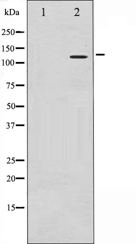
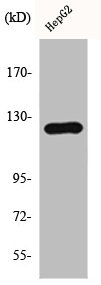
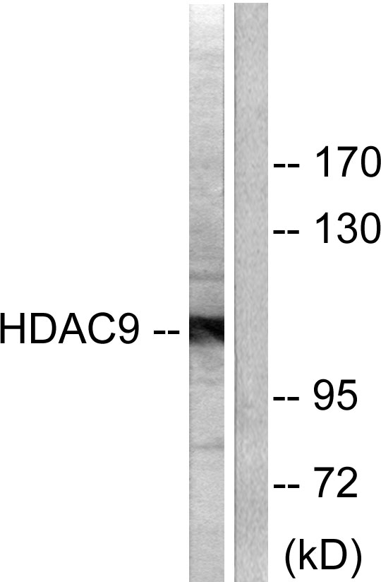
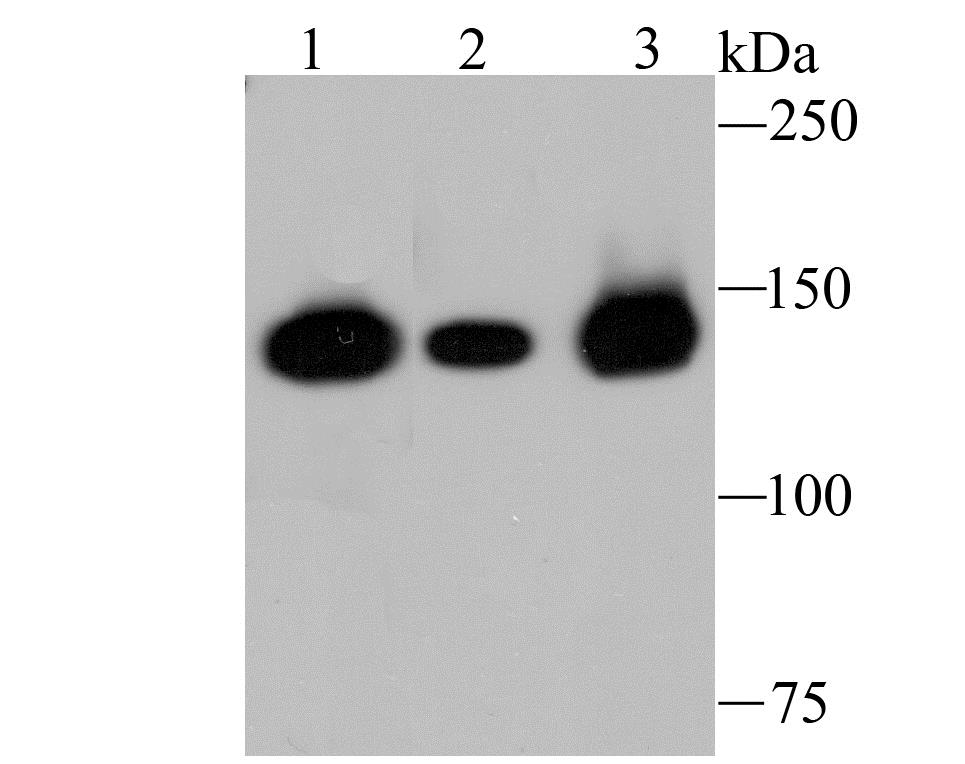
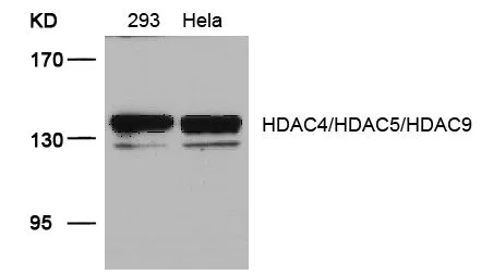
Western blot analysis of HDAC9 expression in HepG2 whole cell lysates,The lane on the left is treated with the antigen-specific peptide.
Western blot analysis of HDAC9 expression in HepG2 whole cell lysates,The lane on the left is treated with the antigen-specific peptide.
HDAC9 antibody
GTX52378
ApplicationsImmunoFluorescence, Western Blot, ImmunoCytoChemistry, ImmunoHistoChemistry
Product group Antibodies
ReactivityHuman
TargetHDAC9
Overview
- SupplierGeneTex
- Product NameHDAC9 antibody
- Delivery Days Customer9
- Application Supplier NoteWB 1:500-1:2000, IHC 1:50-1:200, IF/ICC 1:100-1:500
- ApplicationsImmunoFluorescence, Western Blot, ImmunoCytoChemistry, ImmunoHistoChemistry
- CertificationResearch Use Only
- ClonalityPolyclonal
- Concentration1 mg/ml
- ConjugateUnconjugated
- Gene ID9734
- Target nameHDAC9
- Target descriptionhistone deacetylase 9
- Target synonymsARCND4, HD7, HD7b, HD9, HDAC, HDAC7, HDAC7B, HDAC9B, HDAC9FL, HDRP, MITR, histone deacetylase 9, MEF-2 interacting transcription repressor (MITR) protein, histone deacetylase 4/5-related protein, histone deacetylase 7B
- HostRabbit
- IsotypeIgG
- Protein IDQ9UKV0
- Protein NameHistone deacetylase 9
- Scientific DescriptionHistones play a critical role in transcriptional regulation, cell cycle progression, and developmental events. Histone acetylation/deacetylation alters chromosome structure and affects transcription factor access to DNA. The protein encoded by this gene has sequence homology to members of the histone deacetylase family. This gene is orthologous to the Xenopus and mouse MITR genes. The MITR protein lacks the histone deacetylase catalytic domain. It represses MEF2 activity through recruitment of multicomponent corepressor complexes that include CtBP and HDACs. This encoded protein may play a role in hematopoiesis. Multiple alternatively spliced transcripts have been described for this gene but the full-length nature of some of them has not been determined. [provided by RefSeq, Jul 2008]
- ReactivityHuman
- Storage Instruction-20°C or -80°C,2°C to 8°C
- UNSPSC41116161
Datasheet
Related products
Product group Antibodies
HDAC5/HDAC9 AntibodyCSB-PA002890
ApplicationsWestern Blot, ELISA, ImmunoHistoChemistry
ReactivityHuman, Mouse
TargetHDAC9
- SizePrice

Product group Antibodies
Anti-HDAC9 AntibodyA98657
ApplicationsImmunoFluorescence, Western Blot, ELISA, ImmunoHistoChemistry
ReactivityHuman
- SizePrice

Product group Antibodies
Anti-HDAC9 Antibody Picoband(r)A02177-4-CARRIER-FREE
ApplicationsFlow Cytometry, ImmunoFluorescence, Western Blot, ELISA, ImmunoCytoChemistry
ReactivityHuman, Mouse, Rat
TargetHDAC9
- SizePrice

Product group Antibodies
Anti-HDAC9 AntibodyHPA028926
ApplicationsImmunoCytoChemistry, ImmunoHistoChemistry
ReactivityHuman
TargetHDAC9
- SizePrice
Product group Antibodies

HDAC9 AntibodyLS-C331507
ApplicationsWestern Blot
ReactivityHuman, Mouse, Rat
TargetHDAC9
- SizePrice
Product group Antibodies
ApplicationsImmunoPrecipitation, Western Blot, ImmunoCytoChemistry, ImmunoHistoChemistry
ReactivityRat
TargetHDAC9
- SizePrice
Product group Antibodies
HDAC9 Recombinant AntibodyBSM-54186R
ApplicationsImmunoFluorescence, Western Blot, ImmunoCytoChemistry, ImmunoHistoChemistry, ImmunoHistoChemistry Frozen, ImmunoHistoChemistry Paraffin
ReactivityHuman
TargetHDAC9
- SizePrice
Product group Antibodies
References
HDAC4/HDAC5/HDAC9 antibodyGTX50861
ApplicationsImmunoFluorescence, Western Blot, ImmunoCytoChemistry, ImmunoHistoChemistry, ImmunoHistoChemistry Paraffin
ReactivityHuman
TargetHDAC9
- SizePrice
Product group Antibodies
ApplicationsWestern Blot, ImmunoHistoChemistry, ImmunoHistoChemistry Paraffin
ReactivityHuman
TargetHDAC9
- SizePrice