

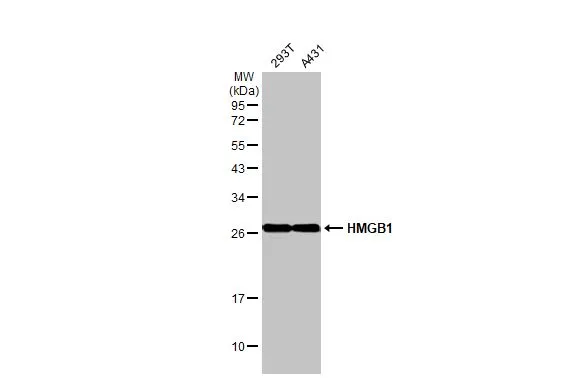
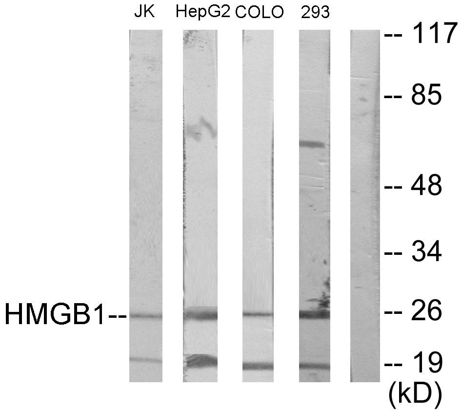
Various whole cell extracts (30 μg) were separated by 12% SDS-PAGE, and the membrane was blotted with HMGB1 antibody [HL2060] (GTX637964) diluted at 1:1000. The HRP-conjugated anti-rabbit IgG antibody (GTX213110-01) was used to detect the primary antibody.
![HMGB1 antibody [HL2060] detects HMGB1 protein at nucleus by immunohistochemical analysis. Sample: Paraffin-embedded mouse tissues. HMGB1 stained by HMGB1 antibody [HL2060] (GTX637964) diluted at 1:100. Antigen Retrieval: Citrate buffer, pH 6.0, 15 min](https://www.genetex.com/upload/website/prouct_img/normal/GTX637964/GTX637964_T-44886_20221230_IHC-P_multiple_M_23020621_195.webp "HMGB1 antibody [HL2060] detects HMGB1 protein at nucleus by immunohistochemical analysis. Sample: Paraffin-embedded mouse tissues. HMGB1 stained by HMGB1 antibody [HL2060] (GTX637964) diluted at 1:100. Antigen Retrieval: Citrate buffer, pH 6.0, 15 min")
![HMGB1 antibody [HL2060] detects HMGB1 protein at nucleus by immunofluorescent analysis. Sample: HeLa cells were fixed in 4% paraformaldehyde at RT for 15 min. Green: HMGB1 stained by HMGB1 antibody [HL2060] (GTX637964) diluted at 1:500. Red: alpha Tubulin, a cytoskeleton marker, stained by alpha Tubulin antibody [GT114] (GTX628802) diluted at 1:1000.](https://www.genetex.com/upload/website/prouct_img/normal/GTX637964/GTX637964_T-44886_20230203_ICC_IF_23021401_442.webp "HMGB1 antibody [HL2060] detects HMGB1 protein at nucleus by immunofluorescent analysis. Sample: HeLa cells were fixed in 4% paraformaldehyde at RT for 15 min. Green: HMGB1 stained by HMGB1 antibody [HL2060] (GTX637964) diluted at 1:500. Red: alpha Tubulin, a cytoskeleton marker, stained by alpha Tubulin antibody [GT114] (GTX628802) diluted at 1:1000.")
![HMGB1 antibody [HL2060] detects HMGB1 protein at nucleus by immunofluorescent analysis. Sample: DIV9 rat E18 primary hippocampal neuron cells were fixed in 4% paraformaldehyde at RT for 15 min. Green: HMGB1 stained by HMGB1 antibody [HL2060] (GTX637964) diluted at 1:250. Red: Tau, an axon marker, stained by Tau antibody [GT287] (GTX634809) diluted at 1:500.](https://www.genetex.com/upload/website/prouct_img/normal/GTX637964/GTX637964_T-44886_20230324_ICC_IF_R_23041023_348.webp "HMGB1 antibody [HL2060] detects HMGB1 protein at nucleus by immunofluorescent analysis. Sample: DIV9 rat E18 primary hippocampal neuron cells were fixed in 4% paraformaldehyde at RT for 15 min. Green: HMGB1 stained by HMGB1 antibody [HL2060] (GTX637964) diluted at 1:250. Red: Tau, an axon marker, stained by Tau antibody [GT287] (GTX634809) diluted at 1:500.")
![HMGB1 antibody [HL2060] detects HMGB1 protein by immunohistochemical analysis. Sample: Paraffin-embedded rat tissue. HMGB1 stained by HMGB1 antibody [HL2060] (GTX637964) diluted at 1:200. Antigen Retrieval: Citrate buffer, pH 6.0, 15 min](https://www.genetex.com/upload/website/prouct_img/normal/GTX637964/GTX637964_44963_20230419_IHC-P_multiple_R_23053001_528.webp "HMGB1 antibody [HL2060] detects HMGB1 protein by immunohistochemical analysis. Sample: Paraffin-embedded rat tissue. HMGB1 stained by HMGB1 antibody [HL2060] (GTX637964) diluted at 1:200. Antigen Retrieval: Citrate buffer, pH 6.0, 15 min")
![Whole cell extract (30 μg) was separated by 12% SDS-PAGE, and the membrane was blotted with HMGB1 antibody [HL2060] (GTX637964) diluted at 1:1000. The HRP-conjugated anti-rabbit IgG antibody (GTX213110-01) was used to detect the primary antibody.](https://www.genetex.com/upload/website/prouct_img/normal/GTX637964/GTX637964_44963_20230616_WB_D_23062019_976.webp "Whole cell extract (30 μg) was separated by 12% SDS-PAGE, and the membrane was blotted with HMGB1 antibody [HL2060] (GTX637964) diluted at 1:1000. The HRP-conjugated anti-rabbit IgG antibody (GTX213110-01) was used to detect the primary antibody.")
![HMGB1 antibody [HL2060] detects HMGB1 protein at cytoplasm and nucleus by immunohistochemical analysis. Sample: Paraffin-embedded cat kidney. HMGB1 stained by HMGB1 antibody [HL2060] (GTX637964) diluted at 1:100. Antigen Retrieval: Citrate buffer, pH 6.0, 15 min](https://www.genetex.com/upload/website/prouct_img/normal/GTX637964/GTX637964_44963_20230721_IHC-P_Cat_23073119_206.webp "HMGB1 antibody [HL2060] detects HMGB1 protein at cytoplasm and nucleus by immunohistochemical analysis. Sample: Paraffin-embedded cat kidney. HMGB1 stained by HMGB1 antibody [HL2060] (GTX637964) diluted at 1:100. Antigen Retrieval: Citrate buffer, pH 6.0, 15 min")
![HMGB1 antibody [HL2060] detects HMGB1 protein by immunofluorescent analysis. Sample: MDCK cells were fixed in 4% paraformaldehyde at RT for 15 min. Green: HMGB1 stained by HMGB1 antibody [HL2060] (GTX637964) diluted at 1:500. Red: alpha Tubulin, a cytoskeleton marker, stained by alpha Tubulin antibody [GT114] (GTX628802) diluted at 1:1000.](https://www.genetex.com/upload/website/prouct_img/normal/GTX637964/GTX637964_44963_20240726_ICC_IF_D_24081300_968.webp "HMGB1 antibody [HL2060] detects HMGB1 protein by immunofluorescent analysis. Sample: MDCK cells were fixed in 4% paraformaldehyde at RT for 15 min. Green: HMGB1 stained by HMGB1 antibody [HL2060] (GTX637964) diluted at 1:500. Red: alpha Tubulin, a cytoskeleton marker, stained by alpha Tubulin antibody [GT114] (GTX628802) diluted at 1:1000.")
![HMGB1 antibody [HL2060] detects HMGB1 protein by immunohistochemical analysis. Sample: Paraffin-embedded human cerebellum. HMGB1 stained by HMGB1 antibody [HL2060] (GTX637964) diluted at 1:400. Antigen Retrieval: Citrate buffer, pH 6.0, 15 min](https://www.genetex.com/upload/website/prouct_img/normal/GTX637964/GTX637964_44963_20251113_IHC-P_2_25112100_913.webp "HMGB1 antibody [HL2060] detects HMGB1 protein by immunohistochemical analysis. Sample: Paraffin-embedded human cerebellum. HMGB1 stained by HMGB1 antibody [HL2060] (GTX637964) diluted at 1:400. Antigen Retrieval: Citrate buffer, pH 6.0, 15 min")
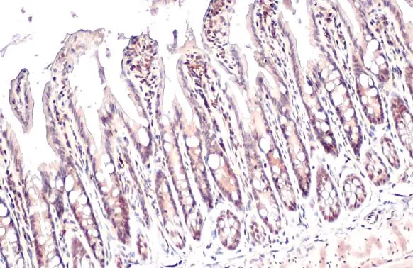
![HMGB1 antibody [HL2060] detects HMGB1 protein by immunohistochemical analysis. Sample: Paraffin-embedded human appendix. HMGB1 stained by HMGB1 antibody [HL2060] (GTX637964) diluted at 1:400. Antigen Retrieval: Citrate buffer, pH 6.0, 15 min](https://www.genetex.com/upload/website/prouct_img/normal/GTX637964/GTX637964_44963_20251113_IHC-P_25112100_326.webp "HMGB1 antibody [HL2060] detects HMGB1 protein by immunohistochemical analysis. Sample: Paraffin-embedded human appendix. HMGB1 stained by HMGB1 antibody [HL2060] (GTX637964) diluted at 1:400. Antigen Retrieval: Citrate buffer, pH 6.0, 15 min")
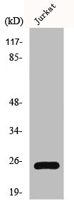
Various whole cell extracts (30 μg) were separated by 12% SDS-PAGE, and the membrane was blotted with HMGB1 antibody [HL2060] (GTX637964) diluted at 1:1000. The HRP-conjugated anti-rabbit IgG antibody (GTX213110-01) was used to detect the primary antibody.
HMGB1 antibody [HL2060]
GTX637964
ApplicationsImmunoFluorescence, Western Blot, ImmunoCytoChemistry, ImmunoHistoChemistry, ImmunoHistoChemistry Paraffin
Product group Antibodies
ReactivityCanine, Feline, Human, Mouse, Rat
TargetHMGB1
Overview
- SupplierGeneTex
- Product NameHMGB1 antibody [HL2060]
- Delivery Days Customer9
- Application Supplier NoteWB: 1:500-1:3000. *Optimal dilutions/concentrations should be determined by the researcher.Not tested in other applications.
- ApplicationsImmunoFluorescence, Western Blot, ImmunoCytoChemistry, ImmunoHistoChemistry, ImmunoHistoChemistry Paraffin
- CertificationResearch Use Only
- ClonalityMonoclonal
- Clone IDHL2060
- Concentration1 mg/ml
- ConjugateUnconjugated
- Gene ID3146
- Target nameHMGB1
- Target descriptionhigh mobility group box 1
- Target synonymsHMG-1, HMG1, HMG3, SBP-1, high mobility group protein B1, Amphoterin, Sulfoglucuronyl carbohydrate binding protein, high-mobility group (nonhistone chromosomal) protein 1
- HostRabbit
- IsotypeIgG
- Protein IDP09429
- Protein NameHigh mobility group protein B1
- Scientific DescriptionThis gene encodes a protein that belongs to the High Mobility Group-box superfamily. The encoded non-histone, nuclear DNA-binding protein regulates transcription, and is involved in organization of DNA. This protein plays a role in several cellular processes, including inflammation, cell differentiation and tumor cell migration. Multiple pseudogenes of this gene have been identified. Alternative splicing results in multiple transcript variants that encode the same protein. [provided by RefSeq, Sep 2015]
- ReactivityCanine, Feline, Human, Mouse, Rat
- Storage Instruction-20°C or -80°C,2°C to 8°C
- UNSPSC41116161
Datasheet
Related products
Product group Antibodies
HMGB1 AntibodyCSB-PA002937
ApplicationsImmunoFluorescence, Western Blot, ELISA, ImmunoHistoChemistry
ReactivityHuman, Mouse, Rat
TargetHMGB1
- SizePrice

Product group Antibodies
Anti-HMGB1 Antibody Picoband(r)A00066-1-CARRIER-FREE
ApplicationsFlow Cytometry, ImmunoFluorescence, Western Blot, ImmunoCytoChemistry, ImmunoHistoChemistry
ReactivityHuman, Mouse, Rat
TargetHMGB1
- SizePrice
Product group Antibodies
Anti-HMGB1 Antibody144-02553
ApplicationsImmunoFluorescence, Western Blot, ImmunoHistoChemistry
ReactivityHuman, Mouse, Rat
TargetHMGB1
- SizePrice
Product group Antibodies
Anti-HMGB1 AntibodyA97486
ApplicationsWestern Blot, ELISA
ReactivityHuman, Mouse, Rat
- SizePrice
Product group Antibodies

HMG1 / HMGB1 AntibodyLS-C766367
ApplicationsImmunoHistoChemistry
ReactivityHuman
TargetHMGB1
- SizePrice

Product group Antibodies
Anti-HMGB1 AntibodyHPA073663
ApplicationsWestern Blot, ImmunoCytoChemistry
ReactivityHuman
TargetHMGB1
- SizePrice
![Various tissue extracts (50 μg) were separated by 12% SDS-PAGE, and the membrane was blotted with HMGB1 antibody [HL2059] (GTX637963) diluted at 1:1000. The HRP-conjugated anti-rabbit IgG antibody (GTX213110-01) was used to detect the primary antibody.](https://www.genetex.com/upload/website/prouct_img/normal/GTX637963/GTX637963_T-44886_20221209_WB_M_R_22121123_430.webp)
Product group Antibodies
HMGB1 antibody [HL2059]GTX637963
ApplicationsImmunoFluorescence, Western Blot, ELISA, ImmunoCytoChemistry, ImmunoHistoChemistry, ImmunoHistoChemistry Paraffin
ReactivityCanine, Feline, Human, Mouse, Rat
TargetHMGB1
- SizePrice
![IHC-P analysis of human placenta (amnion and chorion) tissue using GTX639941 HMGB1 antibody [HMV317] HistoMAX?. Strong nuclear HMGB1 staining of chorion cells and moderate nuclear HMGB1 staining of amnion cells.](https://www.genetex.com/upload/website/prouct_img/normal/GTX639941/GTX639941_20240514_IHC-P_24051319_526.webp)
Product group Antibodies
HMGB1 antibody [HMV317] HistoMAX(tm)GTX639941
ApplicationsImmunoHistoChemistry, ImmunoHistoChemistry Paraffin
ReactivityHuman
TargetHMGB1
- SizePrice
Product group Antibodies
HMGB1 antibodyGTX101277
ApplicationsImmunoFluorescence, Western Blot, ImmunoCytoChemistry, ImmunoHistoChemistry, ImmunoHistoChemistry Paraffin
ReactivityHuman, Mouse, Porcine, Rat, Zebra Fish
TargetHMGB1
- SizePrice