

IgA Secretory Component(SC05), CF405S conjugate, 0.1mg/mL [26628-22-8]
BNC040050
ApplicationsFunctional Assay, Western Blot, ImmunoHistoChemistry, ImmunoHistoChemistry Paraffin
Product group Antibodies
ReactivityBovine, Human, Mouse, Rat
TargetECM1
Overview
- SupplierBiotium
- Product NameIgA Secretory Component(SC05), CF405S conjugate, 0.1mg/mL [26628-22-8]
- Delivery Days Customer9
- ApplicationsFunctional Assay, Western Blot, ImmunoHistoChemistry, ImmunoHistoChemistry Paraffin
- CAS Number26628-22-8
- CertificationResearch Use Only
- ClonalityMonoclonal
- Clone IDSC05
- Concentration0.1 mg/ml
- ConjugateOther Conjugate
- Gene ID1893
- Target nameECM1
- Target descriptionextracellular matrix protein 1
- Target synonymsURBWD, extracellular matrix protein 1, secretory component p85, testicular tissue protein Li 61
- HostMouse
- IsotypeIgG1
- Protein IDQ16610
- Protein NameExtracellular matrix protein 1
- Scientific DescriptionThis MAb reacts with a reduction-resistant epitope present in both free and SIgA bound Secretory Component. It does not react with the cell lines lacking secretory component. The antibody is useful for studying the distribution and level of both free and bound secretory component. Secretory component is differentially expressed in epithelium, and the antibody is a popular marker for identifying subpopulations of epithelial cells and epithelial differentiation. The Secretory component antibody is a useful research tool for studying mucosal immunity, inflammation, remodeling, differentiation and tumorigenesis, all processes associated with differential secretory component expression. Primary antibodies are available purified, or with a selection of fluorescent CF® Dyes and other labels. CF® Dyes offer exceptional brightness and photostability. Note: Conjugates of blue fluorescent dyes like CF®405S and CF®405M are not recommended for detecting low abundance targets, because blue dyes have lower fluorescence and can give higher non-specific background than other dye colors.
- SourceAnimal
- ReactivityBovine, Human, Mouse, Rat
- Storage Instruction2°C to 8°C,RT
- UNSPSC41116161
MSDS
Related products
Product group Antibodies
Anti-ECM1 AntibodyA39010
ApplicationsWestern Blot, ImmunoHistoChemistry
ReactivityHuman
- SizePrice
Product group Antibodies
Anti-ECM1 Antibody144-64315
ApplicationsWestern Blot
ReactivityHuman
TargetECM1
- SizePrice
Product group Antibodies

ECM1 AntibodyLS-C831605
ApplicationsImmunoHistoChemistry
ReactivityHuman, Mouse, Rat
TargetECM1
- SizePrice
Product group Antibodies
References
ECM1 Polyclonal AntibodyBS-0776R
ApplicationsFlow Cytometry, ImmunoFluorescence, Western Blot, ELISA, ImmunoCytoChemistry, ImmunoHistoChemistry, ImmunoHistoChemistry Frozen, ImmunoHistoChemistry Paraffin
ReactivityBovine, Canine, Human, Mouse, Rat
TargetECM1
- SizePrice
Product group Antibodies
ECM1 AntibodyCSB-PA007383LA01HU
ApplicationsWestern Blot, ELISA, ImmunoHistoChemistry
ReactivityHuman
TargetECM1
- SizePrice
Product group Antibodies
ECM1 Polyclonal AntibodyCAC15356
ApplicationsWestern Blot, ELISA, ImmunoHistoChemistry
TargetECM1
- SizePrice

Product group Antibodies
ApplicationsFlow Cytometry, ImmunoFluorescence, ImmunoPrecipitation, Western Blot, ImmunoCytoChemistry, ImmunoHistoChemistry
ReactivityHuman, Mouse, Rat
TargetECM1
- SizePrice
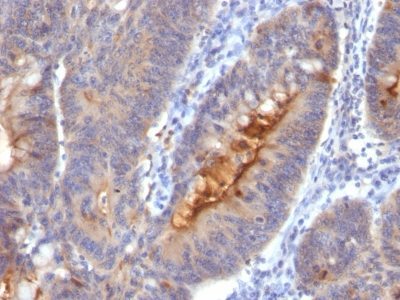
![IHC-P analysis of human colon carcinoma section using GTX02627 ECM1 antibody [ECM1/2889R].](https://www.genetex.com/upload/website/prouct_img/normal/GTX02627/GTX02627_20210319_IHC-P_w_23053122_685.webp)
Product group Antibodies
ECM1 antibody [ECM1/2889R]GTX02627
ApplicationsImmunoHistoChemistry, ImmunoHistoChemistry Paraffin
ReactivityHuman, Rat
TargetECM1
- SizePrice

Product group Antibodies
Anti-ECM1 AntibodyHPA027241
ApplicationsWestern Blot, ImmunoHistoChemistry
ReactivityHuman
TargetECM1
- SizePrice
Product group Antibodies
TargetECM1
- SizePrice