

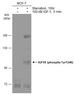
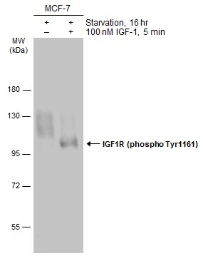
Untreated (–) and treated (+) MCF-7 whole cell extracts (60 μg) were separated by 7.5% SDS-PAGE, and the membrane was blotted with IGF1R beta (phospho Tyr1316) antibodyy (GTX133448) diluted at 1:1000. The HRP-conjugated anti-rabbit IgG antibody (GTX213110-01) was used to detect the primary antibody.
 and treated (+) 293T whole cell extracts (30 μg) were separated by 5% SDS-PAGE, and the membrane was blotted with IGF1R beta (phospho Tyr1316) antibody (GTX133448) diluted at 1:1000. The HRP-conjugated anti-rabbit IgG antibody (GTX213110-01) was used to detect the primary antibody.")
Untreated (–) and treated (+) MCF-7 whole cell extracts (60 μg) were separated by 7.5% SDS-PAGE, and the membrane was blotted with IGF1R beta (phospho Tyr1316) antibodyy (GTX133448) diluted at 1:1000. The HRP-conjugated anti-rabbit IgG antibody (GTX213110-01) was used to detect the primary antibody.
IGF1R beta (phospho Tyr1316) antibody
GTX133448
ApplicationsWestern Blot, ImmunoHistoChemistry, ImmunoHistoChemistry Paraffin
Product group Antibodies
ReactivityHuman
TargetIGF1R
Overview
- SupplierGeneTex
- Product NameIGF1R beta (phospho Tyr1316) antibody
- Delivery Days Customer9
- Application Supplier NoteWB: 1:500-1:3000. *Optimal dilutions/concentrations should be determined by the researcher.Not tested in other applications.
- ApplicationsWestern Blot, ImmunoHistoChemistry, ImmunoHistoChemistry Paraffin
- CertificationResearch Use Only
- ClonalityPolyclonal
- Concentration4 mg/ml
- ConjugateUnconjugated
- Gene ID3480
- Target nameIGF1R
- Target descriptioninsulin like growth factor 1 receptor
- Target synonymsCD221, IGFIR, IGFR, JTK13, insulin-like growth factor 1 receptor, IGF-I receptor
- HostRabbit
- IsotypeIgG
- Protein IDP08069
- Protein NameInsulin-like growth factor 1 receptor
- Scientific DescriptionThis receptor binds insulin-like growth factor with a high affinity. It has tyrosine kinase activity. The insulin-like growth factor I receptor plays a critical role in transformation events. Cleavage of the precursor generates alpha and beta subunits. It is highly overexpressed in most malignant tissues where it functions as an anti-apoptotic agent by enhancing cell survival. [provided by RefSeq]
- ReactivityHuman
- Storage Instruction-20°C or -80°C,2°C to 8°C
- UNSPSC41116161
Datasheet
Related products
Product group Antibodies
Anti-IGF1R AntibodyA95858
ApplicationsWestern Blot, ELISA, ImmunoHistoChemistry
ReactivityHuman, Mouse, Rat
- SizePrice
Product group Antibodies

Anti-IGF-1 [7973 [M23]], Mouse IgG1, kappaAB04263-1.1
ApplicationsWestern Blot, ImmunoHistoChemistry
ReactivityHuman, Mouse, Rat
TargetIGF1R
- SizePrice

Product group Antibodies
Anti-IGF1R Antibody Picoband(r)A00070-3-CARRIER-FREE
ApplicationsWestern Blot, ELISA
ReactivityHuman
TargetIGF1R
- SizePrice

Product group Antibodies
References
IGF1R Polyclonal AntibodyBS-0227R
ApplicationsFlow Cytometry
ReactivityBovine, Canine, Equine, Human, Mouse, Porcine, Rabbit, Rat, Sheep
TargetIGF1R
- SizePrice
Product group Antibodies
IGF1R/INSR AntibodyCSB-PA003003
ApplicationsWestern Blot, ELISA, ImmunoHistoChemistry
ReactivityHuman, Mouse, Rat
TargetIGF1R
- SizePrice
Product group Antibodies
Igf1R Polyclonal AntibodyCAC10405
ApplicationsImmunoFluorescence, ELISA
TargetIGF1R
- SizePrice
Product group Antibodies
IGF1R beta (phospho Tyr950) antibodyGTX133449
ApplicationsWestern Blot
ReactivityHuman
TargetIGF1R
- SizePrice
Product group Antibodies
ApplicationsWestern Blot
ReactivityHuman
TargetIGF1R
- SizePrice
Product group Antibodies
IGF1R beta antibodyGTX134417
ApplicationsWestern Blot
ReactivityHuman
TargetIGF1R
- SizePrice