

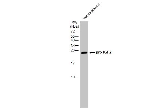
Mouse plasma (30 μg) was separated by 15% SDS-PAGE, and the membrane was blotted with IGF2 antibody [HL1982] (GTX637875) diluted at 1:1000. The HRP-conjugated anti-rabbit IgG antibody (GTX213110-01) was used to detect the primary antibody.
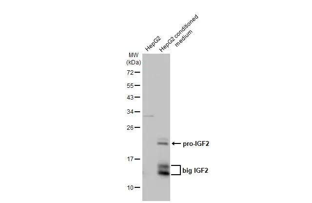
![HepG2 whole cell extract and conditioned medium (30 μg) were separated by 12% SDS-PAGE, and the membrane was blotted with IGF2 antibody [HL1982] (GTX637875) diluted at 1:1000. The HRP-conjugated anti-rabbit IgG antibody (GTX213110-01) was used to detect the primary antibody.](https://www.genetex.com/upload/website/prouct_img/normal/GTX637875/GTX637875_44914_20230113_WB_Fraction_23013122_612.webp "HepG2 whole cell extract and conditioned medium (30 μg) were separated by 12% SDS-PAGE, and the membrane was blotted with IGF2 antibody [HL1982] (GTX637875) diluted at 1:1000. The HRP-conjugated anti-rabbit IgG antibody (GTX213110-01) was used to detect the primary antibody.")
![IGF2 antibody [HL1982] detects IGF2 protein at cytoplasm by immunofluorescent analysis. Sample: HepG2 cells were fixed in 4% paraformaldehyde at RT for 15 min. Green: IGF2 stained by IGF2 antibody [HL1982] (GTX637875) diluted at 1:500. Blue: Fluoroshield with DAPI (GTX30920).](https://www.genetex.com/upload/website/prouct_img/normal/GTX637875/GTX637875_T-44865_20230106_ICC_IF_23013122_935.webp "IGF2 antibody [HL1982] detects IGF2 protein at cytoplasm by immunofluorescent analysis. Sample: HepG2 cells were fixed in 4% paraformaldehyde at RT for 15 min. Green: IGF2 stained by IGF2 antibody [HL1982] (GTX637875) diluted at 1:500. Blue: Fluoroshield with DAPI (GTX30920).")
![Recombinant IGF2 protein were separated by 15% SDS-PAGE, and the membrane was blotted with IGF2 antibody [HL1982] (GTX637875) diluted at 1:2000. The HRP-conjated anti-rabbit IgG antibody (GTX213110-01) was used to detect the primary antibody.](https://www.genetex.com/upload/website/prouct_img/normal/GTX637875/GTX637875_44914_20230414_WB_Ag_23041723_506.webp "Recombinant IGF2 protein were separated by 15% SDS-PAGE, and the membrane was blotted with IGF2 antibody [HL1982] (GTX637875) diluted at 1:2000. The HRP-conjated anti-rabbit IgG antibody (GTX213110-01) was used to detect the primary antibody.")
![Indirect ELISA analysis was performed by coating the plate with recombinant E.coli expressed, mature recombinant human IGF2 protein (533.33-8.33 nM). Coated protein was probed with IGF2 antibody [HL1982] (GTX637875) (1 μg/mL). Goat anti-rabbit IgG antibody (HRP) (GTX213110-01) (1:10000) was used to detect the bound primary antibody.](https://www.genetex.com/upload/website/prouct_img/normal/GTX637875/GTX637875_44914_20230512_ELISA_Indirect_23051702_885.webp "Indirect ELISA analysis was performed by coating the plate with recombinant E.coli expressed, mature recombinant human IGF2 protein (533.33-8.33 nM). Coated protein was probed with IGF2 antibody [HL1982] (GTX637875) (1 μg/mL). Goat anti-rabbit IgG antibody (HRP) (GTX213110-01) (1:10000) was used to detect the bound primary antibody.")
![Non-transfected (–) and transfected (+) 293T whole cell extracts (30 μg) were separated by 15% SDS-PAGE, and the membrane was blotted with IGF2 antibody [HL1982] (GTX637875) diluted at 1:5000. The HRP-conjugated anti-rabbit IgG antibody (GTX213110-01) was used to detect the primary antibody.](https://www.genetex.com/upload/website/prouct_img/normal/GTX637875/GTX637875_44914_20230526_WB_multiple_B_23053001_247.webp "Non-transfected (–) and transfected (+) 293T whole cell extracts (30 μg) were separated by 15% SDS-PAGE, and the membrane was blotted with IGF2 antibody [HL1982] (GTX637875) diluted at 1:5000. The HRP-conjugated anti-rabbit IgG antibody (GTX213110-01) was used to detect the primary antibody.")
![Sandwich ELISA detection of recombinant E.coli expressed, mature recombinant human IGF2 protein using antibodies as below. Capture: IGF2 antibody [HL2937] (GTX640321) (5 μg/mL) Detection: HRP-conjugated IGF2 antibody [HL1982] (GTX637875) (1 μg/mL) Please notice that GTX637875 needs to be conjugated to HRP to function as the detection antibody when paired with GTX640321. Please contact us for custom HRP-conjugated antibody.](https://www.genetex.com/upload/website/prouct_img/normal/GTX637875/GTX637875_44914_20240510_ELISA_PAIR_24051400_773.webp "Sandwich ELISA detection of recombinant E.coli expressed, mature recombinant human IGF2 protein using antibodies as below. Capture: IGF2 antibody [HL2937] (GTX640321) (5 μg/mL) Detection: HRP-conjugated IGF2 antibody [HL1982] (GTX637875) (1 μg/mL) Please notice that GTX637875 needs to be conjugated to HRP to function as the detection antibody when paired with GTX640321. Please contact us for custom HRP-conjugated antibody.")
Mouse plasma (30 μg) was separated by 15% SDS-PAGE, and the membrane was blotted with IGF2 antibody [HL1982] (GTX637875) diluted at 1:1000. The HRP-conjugated anti-rabbit IgG antibody (GTX213110-01) was used to detect the primary antibody.
IGF2 antibody [HL1982]
GTX637875
ApplicationsImmunoFluorescence, Western Blot, ELISA, ImmunoCytoChemistry
Product group Antibodies
ReactivityHuman, Mouse
TargetIGF2
Overview
- SupplierGeneTex
- Product NameIGF2 antibody [HL1982]
- Delivery Days Customer9
- Application Supplier NoteWB: 1:500-1:3000. *Optimal dilutions/concentrations should be determined by the researcher.Not tested in other applications.
- ApplicationsImmunoFluorescence, Western Blot, ELISA, ImmunoCytoChemistry
- CertificationResearch Use Only
- ClonalityMonoclonal
- Clone IDHL1982
- Concentration1 mg/ml
- ConjugateUnconjugated
- Gene ID3481
- Target nameIGF2
- Target descriptioninsulin like growth factor 2
- Target synonymsC11orf43, GRDF, IGF-II, PP9974, SRS3, insulin-like growth factor 2, T3M-11-derived growth factor, insulin-like growth factor 2 (somatomedin A), insulin-like growth factor type 2, preptin
- HostRabbit
- IsotypeIgG
- Protein IDP01344
- Protein NameInsulin-like growth factor 2
- Scientific DescriptionThis gene encodes a member of the insulin family of polypeptide growth factors, which are involved in development and growth. It is an imprinted gene, expressed only from the paternal allele, and epigenetic changes at this locus are associated with Wilms tumour, Beckwith-Wiedemann syndrome, rhabdomyosarcoma, and Silver-Russell syndrome. A read-through INS-IGF2 gene exists, whose 5 region overlaps the INS gene and the 3 region overlaps this gene. Alternatively spliced transcript variants encoding different isoforms have been found for this gene. [provided by RefSeq, Oct 2010]
- ReactivityHuman, Mouse
- Storage Instruction-20°C or -80°C,2°C to 8°C
- UNSPSC12352203
Datasheet
Related products
Product group Antibodies
Anti-IGF2 Antibody144-02086
ApplicationsImmunoFluorescence, Western Blot
ReactivityHuman, Mouse
TargetIGF2
- SizePrice
Product group Antibodies
References
IGF2 antibodyGTX129110
ApplicationsWestern Blot, ImmunoHistoChemistry, ImmunoHistoChemistry Paraffin
ReactivityHuman
TargetIGF2
- SizePrice
![HepG2 whole cell extracts (30 μg) were separated by 12% SDS-PAGE, and the membrane was blotted with IGF2 antibody [N1C3] (GTX100453) diluted at 1:1000. The HRP-conjugated anti-rabbit IgG antibody (GTX213110-01) was used to detect the primary antibody.](https://www.genetex.com/upload/website/prouct_img/normal/GTX100453/GTX100453_44517_20230203_WB_Fraction_23020621_996.webp)
Product group Antibodies
IGF2 antibody [N1C3]GTX100453
ApplicationsImmunoFluorescence, Western Blot, ImmunoCytoChemistry
ReactivityHuman
TargetIGF2
- SizePrice
![WB analysis of HEK293 (1) and IGF2 (AA: 25-180)-hIgGFc transfected HEK293 (2) cell lysate using GTX60630 IGF2 antibody [8H1].](https://www.genetex.com/upload/website/prouct_img/normal/GTX60630/GTX60630_20170912_WB_w_23061123_506.webp)
Product group Antibodies
References
IGF2 antibody [8H1]GTX60630
ApplicationsFlow Cytometry, ImmunoFluorescence, Western Blot, ELISA, ImmunoCytoChemistry, ImmunoHistoChemistry, ImmunoHistoChemistry Paraffin
ReactivityHuman
TargetIGF2
- SizePrice
![Mouse tissue extract (50 μg) was separated by 12% SDS-PAGE, and the membrane was blotted with IGF2 antibody [HL1979] (GTX637872) diluted at 1:1000. The HRP-conjugated anti-rabbit IgG antibody (GTX213110-01) was used to detect the primary antibody.](https://www.genetex.com/upload/website/prouct_img/normal/GTX637872/GTX637872_T-44865_20230127_WB_M_placenta_23013122_169.webp)
Product group Antibodies
IGF2 antibody [HL1979]GTX637872
ApplicationsWestern Blot
ReactivityHuman, Mouse
TargetIGF2
- SizePrice
![HepG2 whole cell extract and conditioned medium (30 μg) were separated by 12% SDS-PAGE, and the membrane was blotted with IGF2 antibody [HL2132] (GTX638104) diluted at 1:1000. The HRP-conjugated anti-rabbit IgG antibody (GTX213110-01) was used to detect the primary antibody.](https://www.genetex.com/upload/website/prouct_img/normal/GTX638104/GTX638104_44998_20230331_WB_Fraction_23041023_266.webp)
Product group Antibodies
IGF2 antibody [HL2132]GTX638104
ApplicationsWestern Blot, ELISA
ReactivityHuman
TargetIGF2
- SizePrice
Product group Antibodies
References
IGF2 Polyclonal AntibodyBS-0015R
ApplicationsFlow Cytometry, ImmunoFluorescence, Western Blot, ELISA, ImmunoCytoChemistry, ImmunoHistoChemistry, ImmunoHistoChemistry Frozen, ImmunoHistoChemistry Paraffin
ReactivityBovine, Canine, Human, Mouse, Porcine, Rabbit, Rat, Sheep
TargetIGF2
- SizePrice