

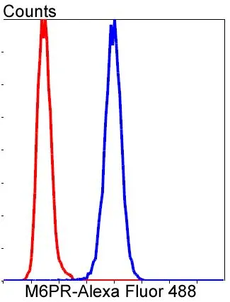
FACS analysis of HeLa cells using GTX64270 IGF2 Receptor antibody [GT1078].
Blue : Primary antibody Red : Isotype control
![ICC/IF analysis of HepG2 cells using GTX64270 IGF2 Receptor antibody [GT1078].
Fixation : 4% PFA
Permeabilization : 0.25% Triton X-100 in PBS
Green : Primary antibody Blue : DAPI](https://www.genetex.com/upload/website/prouct_img/normal/GTX64270/GTX64270_20180608_ICCIF_1_w_23061203_797.webp "ICC/IF analysis of HepG2 cells using GTX64270 IGF2 Receptor antibody [GT1078].
Fixation : 4% PFA
Permeabilization : 0.25% Triton X-100 in PBS
Green : Primary antibody Blue : DAPI")
![ICC/IF analysis of MCF-7 cells using GTX64270 IGF2 Receptor antibody [GT1078].
Fixation : 4% PFA
Permeabilization : 0.25% Triton X-100 in PBS
Green : Primary antibody Blue : DAPI](https://www.genetex.com/upload/website/prouct_img/normal/GTX64270/GTX64270_20180608_ICCIF_2_w_23061203_523.webp "ICC/IF analysis of MCF-7 cells using GTX64270 IGF2 Receptor antibody [GT1078].
Fixation : 4% PFA
Permeabilization : 0.25% Triton X-100 in PBS
Green : Primary antibody Blue : DAPI")
![IHC-P analysis of human tonsil tissue using GTX64270 IGF2 Receptor antibody [GT1078].](https://www.genetex.com/upload/website/prouct_img/normal/GTX64270/GTX64270_20180608_IHC-P_w_23061203_497.webp "IHC-P analysis of human tonsil tissue using GTX64270 IGF2 Receptor antibody [GT1078].")
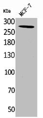
![Various whole cell extracts (30 μg) were separated by 5% SDS-PAGE, and the membrane was blotted with IGF2R antibody [GT1078] (GTX64270) diluted at 1:500. The HRP-conjugated anti-rabbit IgG antibody (GTX213110-01) was used to detect the primary antibody. Corresponding RNA expression data for the same cell lines are based on Human Protein Atlas program.](https://www.genetex.com/upload/website/prouct_img/normal/GTX64270/GTX64270_HM1008_20200228_WB_TPM_watermark_w_23061203_273.webp "Various whole cell extracts (30 μg) were separated by 5% SDS-PAGE, and the membrane was blotted with IGF2R antibody [GT1078] (GTX64270) diluted at 1:500. The HRP-conjugated anti-rabbit IgG antibody (GTX213110-01) was used to detect the primary antibody. Corresponding RNA expression data for the same cell lines are based on Human Protein Atlas program.")
![ICC/IF analysis of HeLa cells using GTX64270 IGF2 Receptor antibody [GT1078].
Fixation : 4% PFA
Permeabilization : 0.25% Triton X-100 in PBS
Green : Primary antibody Blue : DAPI](https://www.genetex.com/upload/website/prouct_img/normal/GTX64270/GTX64270_20180608_ICCIF_w_23061203_834.webp "ICC/IF analysis of HeLa cells using GTX64270 IGF2 Receptor antibody [GT1078].
Fixation : 4% PFA
Permeabilization : 0.25% Triton X-100 in PBS
Green : Primary antibody Blue : DAPI")
FACS analysis of HeLa cells using GTX64270 IGF2 Receptor antibody [GT1078].
Blue : Primary antibody Red : Isotype control
IGF2R antibody [GT1078]
GTX64270
ApplicationsFlow Cytometry, ImmunoFluorescence, Western Blot, ImmunoCytoChemistry, ImmunoHistoChemistry, ImmunoHistoChemistry Paraffin
Product group Antibodies
ReactivityHuman
TargetIGF2R
Overview
- SupplierGeneTex
- Product NameIGF2R antibody [GT1078]
- Delivery Days Customer9
- Application Supplier NoteWB: 1:1000-5000. ICC/IF: 1:50-1:200. IHC-P: 1:50-1:200. FACS: 1:50-1:100. *Optimal dilutions/concentrations should be determined by the researcher.Not tested in other applications.
- ApplicationsFlow Cytometry, ImmunoFluorescence, Western Blot, ImmunoCytoChemistry, ImmunoHistoChemistry, ImmunoHistoChemistry Paraffin
- CertificationResearch Use Only
- ClonalityMonoclonal
- Clone IDGT1078
- ConjugateUnconjugated
- Gene ID3482
- Target nameIGF2R
- Target descriptioninsulin like growth factor 2 receptor
- Target synonymsCD222, CI-M6PR, CIMPR, M6P-R, M6P/IGF2R, MPR 300, MPR1, MPR300, MPRI, cation-independent mannose-6-phosphate receptor, 300 kDa mannose 6-phosphate receptor, CI Man-6-P receptor, IGF-II receptor, M6P/IGF2 receptor, insulin-like growth factor II receptor
- HostRabbit
- IsotypeIgG
- Scientific DescriptionThis gene encodes a receptor for both insulin-like growth factor 2 and mannose 6-phosphate, although the binding sites for either are located on different segments of the receptor. This receptor functions in the intracellular trafficking of lysosomal enzymes, the activation of transforming growth factor beta, and the degradation of insulin-like growth factor 2. While the related mouse gene shows exclusive expression from the maternal allele, imprinting of the human gene appears to be polymorphic, with only a minority of individuals showing expression from the maternal allele. [provided by RefSeq, Apr 2013]
- ReactivityHuman
- Storage Instruction-20°C or -80°C,2°C to 8°C
- UNSPSC12352203
Datasheet
Related products
Product group Antibodies

IGF2R (Phospho-Ser2409) AntibodyABX012519
ApplicationsWestern Blot, ELISA, ImmunoHistoChemistry
- SizePrice
Product group Antibodies
Anti-IGF2R Antibody144-60151
ApplicationsImmunoFluorescence, Western Blot, ImmunoHistoChemistry
ReactivityHuman, Mouse, Rat
TargetIGF2R
- SizePrice
Product group Antibodies
ApplicationsImmunoPrecipitation, Western Blot, ImmunoCytoChemistry, ImmunoHistoChemistry
TargetIGF2R
- SizePrice
Product group Antibodies
IGF2R/M6PR Polyclonal AntibodyBS-6670R
ApplicationsFlow Cytometry, ImmunoFluorescence, ELISA, ImmunoCytoChemistry, ImmunoHistoChemistry, ImmunoHistoChemistry Frozen, ImmunoHistoChemistry Paraffin
ReactivityBovine, Canine, Chicken, Equine, Human, Mouse, Porcine, Rabbit, Rat
TargetIGF2R
- SizePrice
Product group Antibodies
IGF2R AntibodyCSB-PA005283
ApplicationsWestern Blot, ELISA
ReactivityHuman
TargetIGF2R
- SizePrice

Product group Antibodies
Anti-Mannose 6 Phosphate Receptor (Cation independent)/IGF2R Antibody Picoband(r)A00951-CARRIER-FREE
ApplicationsFlow Cytometry, ImmunoFluorescence, Western Blot, ELISA, ImmunoCytoChemistry, ImmunoHistoChemistry
ReactivityHuman, Mouse, Rat
TargetIGF2R
- SizePrice
Product group Antibodies
ReactivityHuman
TargetIGF2R
- SizePrice

Product group Antibodies
Anti-IGF2R AntibodyHPA011332
ApplicationsImmunoCytoChemistry, ImmunoHistoChemistry
ReactivityHuman
TargetIGF2R
- SizePrice
Product group Antibodies
References
IGF2R antibody [MEM-238] (Biotin)GTX28093-02
ApplicationsFlow Cytometry, ImmunoPrecipitation, Western Blot
ReactivityHuman, Primate
TargetIGF2R
- SizePrice