

Mouse tissue extract (50 μg) was separated by 15% SDS-PAGE, and the membrane was blotted with Insulin antibody [GT1229] (GTX02826) diluted at 1:1000. The HRP-conjugated anti-rabbit IgG antibody (GTX213110-01) was used to detect the primary antibody.
![WB analysis of various samples using GTX02826 Insulin antibody [GT1229]. Dilution : 1:1000 Loading : 25μg](https://www.genetex.com/upload/website/prouct_img/normal/GTX02826/CutImage_A19066_WB_01_(1068937)_w_23053123_191.webp "WB analysis of various samples using GTX02826 Insulin antibody [GT1229]. Dilution : 1:1000 Loading : 25μg")
![IHC-P analysis of rat pancreas tissue section using Insulin antibody [GT1229] Insulin antibody [GT1229]. Dilution : 1:100 Blue : DAPI for nuclear staining.](https://www.genetex.com/upload/website/prouct_img/normal/GTX02826/A19066_IF_01_(1068664)_w_23053123_316.webp "IHC-P analysis of rat pancreas tissue section using Insulin antibody [GT1229] Insulin antibody [GT1229]. Dilution : 1:100 Blue : DAPI for nuclear staining.")
![Insulin antibody [GT1229] detects Insulin protein at cytoplasm by immunohistochemical analysis. Sample: Paraffin-embedded rat pancreas. Insulin stained by Insulin antibody [GT1229] (GTX02826) diluted at 1:500. Antigen Retrieval: Citrate buffer, pH 6.0, 15 min](https://www.genetex.com/upload/website/prouct_img/normal/GTX02826/GTX02826_20210129_IHC-P_R_w_23053123_275.webp "Insulin antibody [GT1229] detects Insulin protein at cytoplasm by immunohistochemical analysis. Sample: Paraffin-embedded rat pancreas. Insulin stained by Insulin antibody [GT1229] (GTX02826) diluted at 1:500. Antigen Retrieval: Citrate buffer, pH 6.0, 15 min")
![Insulin antibody [GT1229] detects Insulin protein at cytoplasm by immunohistochemical analysis. Sample: Paraffin-embedded mouse pancreas. Insulin stained by Insulin antibody [GT1229] (GTX02826) diluted at 1:500. Antigen Retrieval: Citrate buffer, pH 6.0, 15 min](https://www.genetex.com/upload/website/prouct_img/normal/GTX02826/GTX02826_20210129_IHC-P_M_w_23053123_849.webp "Insulin antibody [GT1229] detects Insulin protein at cytoplasm by immunohistochemical analysis. Sample: Paraffin-embedded mouse pancreas. Insulin stained by Insulin antibody [GT1229] (GTX02826) diluted at 1:500. Antigen Retrieval: Citrate buffer, pH 6.0, 15 min")
![IHC-P analysis of mouse pancreas tissue section using Insulin antibody [GT1229] Insulin antibody [GT1229]. Dilution : 1:100 Blue : DAPI for nuclear staining.](https://www.genetex.com/upload/website/prouct_img/normal/GTX02826/A19066_IF_02_(1068663)_w_23053123_720.webp "IHC-P analysis of mouse pancreas tissue section using Insulin antibody [GT1229] Insulin antibody [GT1229]. Dilution : 1:100 Blue : DAPI for nuclear staining.")
![Rat tissue extract (50 μg) was separated by 15% SDS-PAGE, and the membrane was blotted with Insulin antibody [GT1229] (GTX02826) diluted at 1:1000. The HRP-conjugated anti-rabbit IgG antibody (GTX213110-01) was used to detect the primary antibody.](https://www.genetex.com/upload/website/prouct_img/normal/GTX02826/GTX02826_4000000209_20210122_WB_R_pancreas_w_23053123_778.webp "Rat tissue extract (50 μg) was separated by 15% SDS-PAGE, and the membrane was blotted with Insulin antibody [GT1229] (GTX02826) diluted at 1:1000. The HRP-conjugated anti-rabbit IgG antibody (GTX213110-01) was used to detect the primary antibody.")
![IHC-P analysis of rat pancreas tissue (pancreatic islet) using GTX02826 Insulin antibody [GT1229]. Dilution : 1:100](https://www.genetex.com/upload/website/prouct_img/normal/GTX02826/A19066_IHC_01_(1068592)_w_23053123_307.webp "IHC-P analysis of rat pancreas tissue (pancreatic islet) using GTX02826 Insulin antibody [GT1229]. Dilution : 1:100")
![IHC-P analysis of mouse pancreas tissue (pancreatic islet) using GTX02826 Insulin antibody [GT1229]. Dilution : 1:100](https://www.genetex.com/upload/website/prouct_img/normal/GTX02826/A19066_IHC_02_(1068593)_w_23053123_210.webp "IHC-P analysis of mouse pancreas tissue (pancreatic islet) using GTX02826 Insulin antibody [GT1229]. Dilution : 1:100")
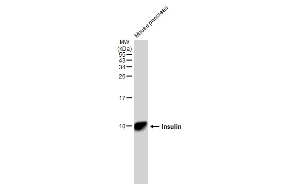
Mouse tissue extract (50 μg) was separated by 15% SDS-PAGE, and the membrane was blotted with Insulin antibody [GT1229] (GTX02826) diluted at 1:1000. The HRP-conjugated anti-rabbit IgG antibody (GTX213110-01) was used to detect the primary antibody.
Insulin antibody [GT1229]
GTX02826
ApplicationsWestern Blot, ImmunoHistoChemistry, ImmunoHistoChemistry Paraffin
Product group Antibodies
ReactivityHuman, Mouse, Rat
TargetINS
Overview
- SupplierGeneTex
- Product NameInsulin antibody [GT1229]
- Delivery Days Customer9
- Application Supplier NoteWB: 1:500 - 1:2000. IHC-P: 1:50 - 1:200. *Optimal dilutions/concentrations should be determined by the researcher.Not tested in other applications.
- ApplicationsWestern Blot, ImmunoHistoChemistry, ImmunoHistoChemistry Paraffin
- CertificationResearch Use Only
- ClonalityMonoclonal
- Clone IDGT1229
- Concentration0.61 mg/ml
- ConjugateUnconjugated
- Gene ID3630
- Target nameINS
- Target descriptioninsulin
- Target synonymsIDDM, IDDM1, IDDM2, ILPR, IRDN, MODY10, PNDM4, insulin, preproinsulin, proinsulin
- HostRabbit
- IsotypeIgG
- Protein IDP01308
- Protein NameInsulin
- Scientific DescriptionAfter removal of the precursor signal peptide, proinsulin is post-translationally cleaved into three peptides: the B chain and A chain peptides, which are covalently linked via two disulfide bonds to form insulin, and C-peptide. Binding of insulin to the insulin receptor (INSR) stimulates glucose uptake. A multitude of mutant alleles with phenotypic effects have been identified. There is a read-through gene, INS-IGF2, which overlaps with this gene at the 5 region and with the IGF2 gene at the 3 region. Alternative splicing results in multiple transcript variants. [provided by RefSeq, Jun 2010]
- ReactivityHuman, Mouse, Rat
- Storage Instruction-20°C,2°C to 8°C
- UNSPSC12352203
Datasheet
Related products
Product group Antibodies

Anti-Insulin [HB125 (mAb1, AE9D6, Ab 125)]Ab03131-1.1
ApplicationsImmunoFluorescence, ELISA, ImmunoHistoChemistry
ReactivityHuman, Porcine, Rodent
TargetINS
- SizePrice
Product group Antibodies
Anti-Insulin Antibody130-11014
ApplicationsELISA
ReactivityHuman
TargetINS
- SizePrice

Product group Antibodies
References
ApplicationsImmunoFluorescence, ImmunoCytoChemistry, ImmunoHistoChemistry
ReactivityHuman, Mouse, Rat
TargetINS
- SizePrice

Product group Antibodies
ApplicationsELISA
ReactivityHuman
TargetINS
- SizePrice
Product group Antibodies
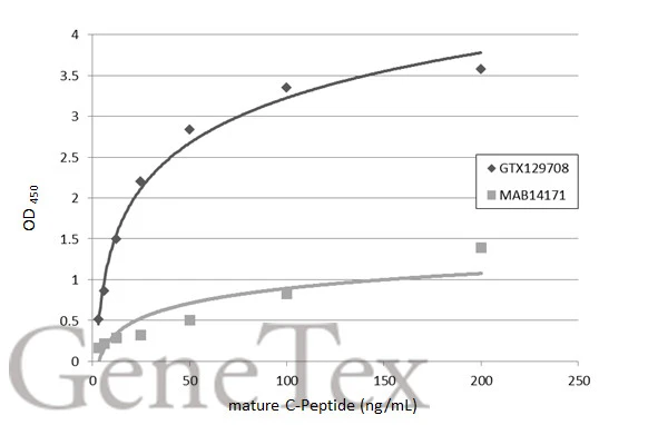
C-Peptide antibodyGTX129708
ApplicationsELISA, ImmunoHistoChemistry, ImmunoHistoChemistry Paraffin
ReactivityHuman, Mouse, Rat
TargetINS
- SizePrice
Product group Antibodies
C-Peptide antibody [GT1455]GTX631941
ApplicationsELISA
ReactivityHuman
TargetINS
- SizePrice
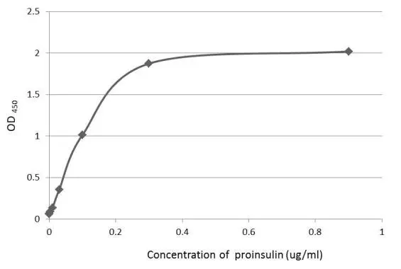
![An ELISA plate is coated with 50 μL of a Proinsulin recombinant protein at concentration ranged from 0.2 μg/mL to 1.6 μg/mL. The coated protein is detected with anti-C-peptide antibody [GT1489] (GTX631942) at 500 ng/mL.](https://www.genetex.com/upload/website/prouct_img/normal/GTX631942/GTX631942_41848_20150206_ELISA_w_23061202_525.webp)
Product group Antibodies
C-Peptide antibody [GT1489]GTX631942
ApplicationsELISA
ReactivityHuman
TargetINS
- SizePrice
![An ELISA plate is coated with 50 μL of a Proinsulin recombinant protein at concentration ranged from 0.2 μg/mL to 1.6 μg/mL. The coated protein is detected with anti-C-peptide antibody [GT1519] (GTX631943) at 500 ng/mL.](https://www.genetex.com/upload/website/prouct_img/normal/GTX631943/GTX631943_41848_20150206_ELISA_w_23061202_907.webp)
Product group Antibodies
C-Peptide antibody [GT1519]GTX631943
ApplicationsELISA
ReactivityHuman
TargetINS
- SizePrice
![Indirect ELISA analysis performed by coating plate with recombinant mature C-Peptide protein (19.05-0.3 nM). Coated protein was probed with C-Peptide antibody [HL1158] (GTX636462) (1 μg/mL). Goat anti-rabbit IgG antibody (HRP) (GTX213110-01) (1:10000) was used to detect bound primary antibody.](https://www.genetex.com/upload/website/prouct_img/normal/GTX636462/GTX636462_44466_20211105_ELISA_Indirect_w_23061202_827.webp)
Product group Antibodies
C-Peptide antibody [HL1158]GTX636462
ApplicationsELISA, ImmunoHistoChemistry, ImmunoHistoChemistry Paraffin
ReactivityHuman, Mouse, Rat
TargetINS
- SizePrice