

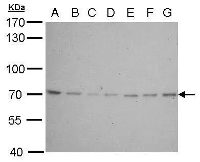
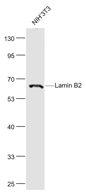
Lamin B2 antibody [N3C2], Internal detects LMNB2 protein by western blot analysis. A. 30 μg Neuro2A whole cell lysate/extract B. 30 μg GL261 whole cell lysate/extract C. 30 μg C8D30 whole cell lysate/extract D. 30 μg NIH-3T3 whole cell lysate/extract E. 30 μg BCL-1 lysate/extract F. 30 μg Raw264.7 whole cell lysate/extract G. 30 μg C2C12 whole cell lysate/extract 7.5% SDS-PAGE Lamin B2 antibody [N3C2], Internal (GTX109894) dilution: 1:500 The HRP-conjugated anti-rabbit IgG antibody (GTX213110-01) was used to detect the primary antibody.
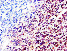
![Lamin B2 antibody [N3C2], Internal detects LMNB2 protein at on Cal27 xenograft by immunohistochemical analysis. Sample: Paraffin-embedded Cal27 xenograft. Lamin B2 antibody [N3C2], Internal (GTX109894) dilution: 1:500.
Antigen Retrieval: Trilogy? (EDTA based, pH 8.0) buffer, 15min](https://www.genetex.com/upload/website/prouct_img/normal/GTX109894/GTX109894_40079_IHC_w_23060500_416.webp "Lamin B2 antibody [N3C2], Internal detects LMNB2 protein at on Cal27 xenograft by immunohistochemical analysis. Sample: Paraffin-embedded Cal27 xenograft. Lamin B2 antibody [N3C2], Internal (GTX109894) dilution: 1:500.
Antigen Retrieval: Trilogy? (EDTA based, pH 8.0) buffer, 15min")
![Lamin B2 antibody [N3C2], Internal immunoprecipitates Lamin B2 protein in IP experiments. IP Sample: HepG2 whole cell lysate/extract A : 30 μg whole cell lysate/extract of Lamin B2 protein expressing HepG2 cells B : Control with 3 μg of pre-immune rabbit IgG C : Immunoprecipitation of Lamin B2 by 3 μg of Lamin B2 antibody [N3C2], Internal (GTX109894) 7.5% SDS-PAGE The immunoprecipitated Lamin B2 protein was detected by Lamin B2 antibody [N3C2], Internal (GTX109894) diluted at 1 : 1000. EasyBlot anti-rabbit IgG (HRP) (GTX221666-01) was used as a secondary reagent.](https://www.genetex.com/upload/website/prouct_img/normal/GTX109894/GTX109894_40079_IP_w_23060500_475.webp "Lamin B2 antibody [N3C2], Internal immunoprecipitates Lamin B2 protein in IP experiments. IP Sample: HepG2 whole cell lysate/extract A : 30 μg whole cell lysate/extract of Lamin B2 protein expressing HepG2 cells B : Control with 3 μg of pre-immune rabbit IgG C : Immunoprecipitation of Lamin B2 by 3 μg of Lamin B2 antibody [N3C2], Internal (GTX109894) 7.5% SDS-PAGE The immunoprecipitated Lamin B2 protein was detected by Lamin B2 antibody [N3C2], Internal (GTX109894) diluted at 1 : 1000. EasyBlot anti-rabbit IgG (HRP) (GTX221666-01) was used as a secondary reagent.")
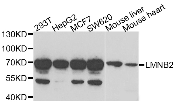
![Various whole cell extracts (30 μg) were separated by 7.5% SDS-PAGE, and the membrane was blotted with Lamin B2 antibody [N3C2], Internal (GTX109894) diluted at 1:1000. The HRP-conjugated anti-rabbit IgG antibody (GTX213110-01) was used to detect the primary antibody.](https://www.genetex.com/upload/website/prouct_img/normal/GTX109894/GTX109894_45364_20240329_WB_24041019_720.webp "Various whole cell extracts (30 μg) were separated by 7.5% SDS-PAGE, and the membrane was blotted with Lamin B2 antibody [N3C2], Internal (GTX109894) diluted at 1:1000. The HRP-conjugated anti-rabbit IgG antibody (GTX213110-01) was used to detect the primary antibody.")
Lamin B2 antibody [N3C2], Internal detects LMNB2 protein by western blot analysis. A. 30 μg Neuro2A whole cell lysate/extract B. 30 μg GL261 whole cell lysate/extract C. 30 μg C8D30 whole cell lysate/extract D. 30 μg NIH-3T3 whole cell lysate/extract E. 30 μg BCL-1 lysate/extract F. 30 μg Raw264.7 whole cell lysate/extract G. 30 μg C2C12 whole cell lysate/extract 7.5% SDS-PAGE Lamin B2 antibody [N3C2], Internal (GTX109894) dilution: 1:500 The HRP-conjugated anti-rabbit IgG antibody (GTX213110-01) was used to detect the primary antibody.
Lamin B2 antibody [N3C2], Internal
GTX109894
ApplicationsImmunoPrecipitation, Western Blot, ImmunoHistoChemistry, ImmunoHistoChemistry Paraffin
Product group Antibodies
ReactivityHuman, Mouse
TargetLMNB2
Overview
- SupplierGeneTex
- Product NameLamin B2 antibody [N3C2], Internal
- Delivery Days Customer9
- Application Supplier NoteWB: 1:500-1:40000. IHC-P: 1:100-1:1000. IP: 1:100-1:500. *Optimal dilutions/concentrations should be determined by the researcher.Not tested in other applications.
- ApplicationsImmunoPrecipitation, Western Blot, ImmunoHistoChemistry, ImmunoHistoChemistry Paraffin
- CertificationResearch Use Only
- ClonalityPolyclonal
- Concentration0.58 mg/ml
- ConjugateUnconjugated
- Gene ID84823
- Target nameLMNB2
- Target descriptionlamin B2
- Target synonymsEPM9, LAMB2, LMN2, MCPH27, lamin-B2, epididymis secretory sperm binding protein, lamin B3
- HostRabbit
- IsotypeIgG
- Protein IDQ03252
- Protein NameLamin-B2
- Scientific DescriptionThe nuclear lamina consists of a two-dimensional matrix of proteins located next to the inner nuclear membrane. The lamin family of proteins make up the matrix and are highly conserved in evolution. During mitosis, the lamina matrix is reversibly disassembled as the lamin proteins are phosphorylated. Lamin proteins are thought to be involved in nuclear stability, chromatin structure and gene expression. Vertebrate lamins consist of two types, A and B. This gene encodes one of the two B type proteins, B2. This gene is in a head-to-tail orientation with the gene for the translocase of inner mitochondrial membrane 13 homolog gene. [provided by RefSeq]
- ReactivityHuman, Mouse
- Storage Instruction-20°C or -80°C,2°C to 8°C
- UNSPSC41116161
Datasheet
Related products

Product group Antibodies
Anti-Lamin B2/LMNB2 Antibody Picoband(r)A05348-1-CARRIER-FREE
ApplicationsWestern Blot, ELISA, ImmunoHistoChemistry
ReactivityHuman, Rat
TargetLMNB2
- SizePrice
Product group Antibodies
Anti-LMNB2 AntibodyA31420
ApplicationsImmunoFluorescence, Western Blot, ImmunoHistoChemistry
ReactivityHuman, Mouse, Rat
- SizePrice
Product group Antibodies
Anti-LMNB2 Antibody144-06483
ApplicationsImmunoFluorescence, Western Blot, ImmunoHistoChemistry
ReactivityHuman, Mouse, Rat
TargetLMNB2
- SizePrice
Product group Antibodies
Lamin B2 Polyclonal AntibodyBS-11132R
ApplicationsImmunoFluorescence, Western Blot, ELISA, ImmunoCytoChemistry, ImmunoHistoChemistry, ImmunoHistoChemistry Frozen, ImmunoHistoChemistry Paraffin
TargetLMNB2
- SizePrice
Product group Antibodies
LMNB2 AntibodyCSB-PA013005LA01HU
ApplicationsImmunoFluorescence, ELISA, ImmunoHistoChemistry
ReactivityHuman
TargetLMNB2
- SizePrice
Product group Antibodies

Mouse anti Lamin B2MUB1104P-CE/IVD
ApplicationsFlow Cytometry, Western Blot, ImmunoHistoChemistry, ImmunoHistoChemistry Frozen
ReactivityHamster, Human, Mouse, Porcine, Xenopus, Zebra Fish
TargetLMNB2
- SizePrice
Product group Antibodies

LMNB2 / Lamin B2 AntibodyLS-C482627
ApplicationsImmunoFluorescence, Western Blot, ImmunoCytoChemistry, ImmunoHistoChemistry, ImmunoHistoChemistry Paraffin
ReactivityHuman, Mouse, Rat
TargetLMNB2
- SizePrice
![Lane 1: Marker [kDa] 250, 130, 95, 72, 55, 36, 28, 17, 10. Lane 2: Human cell line RT-4. Lane 3: Human cell line U-251 MG. Lane 4: Human plasma. Lane 5: Human Liver tissue. Lane 6: Human Tonsil tissue](https://atlasantibodies.s3.amazonaws.com/images/wb/hpa047863-wb-1.jpg)
Product group Antibodies
Anti-LMNB2 AntibodyHPA047863
ApplicationsWestern Blot, ImmunoHistoChemistry
ReactivityHuman
TargetLMNB2
- SizePrice
Product group Antibodies
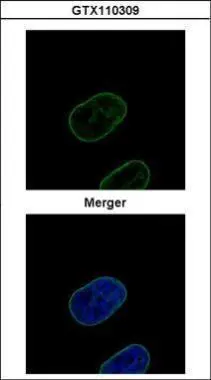
Lamin B2 antibodyGTX110309
ApplicationsImmunoFluorescence, Western Blot, ImmunoCytoChemistry, ImmunoHistoChemistry, ImmunoHistoChemistry Paraffin
ReactivityHuman
TargetLMNB2
- SizePrice
Product group Antibodies
Lamin B2 antibody [GT144]GTX628803
ApplicationsImmunoFluorescence, ImmunoPrecipitation, Western Blot, ImmunoCytoChemistry, ImmunoHistoChemistry, ImmunoHistoChemistry Paraffin
ReactivityHuman
TargetLMNB2
- SizePrice