

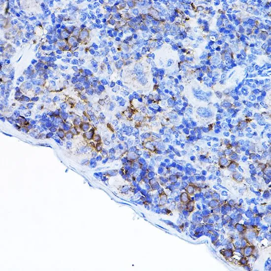
IHC-P analysis of mouse spleen tissue using GTX54287 Lck antibody. Dilution : 1:100


IHC-P analysis of mouse spleen tissue using GTX54287 Lck antibody. Dilution : 1:100
Lck antibody
GTX54287
ApplicationsWestern Blot, ImmunoHistoChemistry, ImmunoHistoChemistry Paraffin
Product group Antibodies
ReactivityHuman, Mouse, Rat
TargetLCK
Overview
- SupplierGeneTex
- Product NameLck antibody
- Delivery Days Customer7
- Application Supplier NoteWB: 1:500 - 1:2000. IHC-P: 1:50 - 1:200. *Optimal dilutions/concentrations should be determined by the researcher.Not tested in other applications.
- ApplicationsWestern Blot, ImmunoHistoChemistry, ImmunoHistoChemistry Paraffin
- CertificationResearch Use Only
- ClonalityPolyclonal
- ConjugateUnconjugated
- Gene ID3932
- Target nameLCK
- Target descriptionLCK proto-oncogene, Src family tyrosine kinase
- Target synonymsIMD22, LSK, YT16, p56lck, pp58lck, tyrosine-protein kinase Lck, T-lymphocyte specific protein tyrosine kinase p56lck, leukocyte C-terminal Src kinase, lymphocyte cell-specific protein-tyrosine kinase, p56(LSTRA) protein-tyrosine kinase, proto-oncogene tyrosine-protein kinase LCK, t cell-specific protein-tyrosine kinase
- HostRabbit
- IsotypeIgG
- Protein IDP06239
- Protein NameTyrosine-protein kinase Lck
- Scientific DescriptionThis gene is a member of the Src family of protein tyrosine kinases (PTKs). The encoded protein is a key signaling molecule in the selection and maturation of developing T-cells. It contains N-terminal sites for myristylation and palmitylation, a PTK domain, and SH2 and SH3 domains which are involved in mediating protein-protein interactions with phosphotyrosine-containing and proline-rich motifs, respectively. The protein localizes to the plasma membrane and pericentrosomal vesicles, and binds to cell surface receptors, including CD4 and CD8, and other signaling molecules. Multiple alternatively spliced variants encoding different isoforms have been described. [provided by RefSeq, Aug 2016]
- ReactivityHuman, Mouse, Rat
- Storage Instruction-20°C or -80°C,2°C to 8°C
- UNSPSC41116161
Datasheet
Related products
Product group Antibodies
Anti-Lck AntibodyA96648
ApplicationsImmunoFluorescence, Western Blot, ELISA
ReactivityHuman, Mouse, Rat
- SizePrice
Product group Antibodies
Anti-LCK Antibody144-02177
ApplicationsWestern Blot
ReactivityHuman, Mouse
TargetLCK
- SizePrice
Product group Antibodies
LCK Recombinant Antibody, Biotin ConjugatedBSM-61647R-BIOTIN
ApplicationsImmunoPrecipitation, Western Blot, ImmunoHistoChemistry, ImmunoHistoChemistry Frozen, ImmunoHistoChemistry Paraffin
ReactivityHuman
TargetLCK
- SizePrice
Product group Antibodies
LCK AntibodyCSB-PA003142
ApplicationsWestern Blot, ELISA
ReactivityHuman, Mouse, Rat
TargetLCK
- SizePrice
Product group Antibodies

Goat anti-LCK (aa39-52)EB11387
ApplicationsWestern Blot, ELISA
ReactivityBovine, Canine, Human, Mouse, Porcine, Rat
TargetLCK
- SizePrice
Product group Antibodies
LCK Polyclonal AntibodyCAC13798
ApplicationsImmunoFluorescence, ImmunoPrecipitation, ELISA, ImmunoHistoChemistry
TargetLCK
- SizePrice
Product group Antibodies
Lck (phospho Tyr394) antibodyGTX133876
ApplicationsWestern Blot
ReactivityHuman, Mouse, Rat
TargetLCK
- SizePrice
Product group Antibodies
Lck antibodyGTX13953
ApplicationsWestern Blot
ReactivityHuman, Mouse
TargetLCK
- SizePrice
Product group Antibodies
Lck (phospho Tyr505) antibodyGTX24901
ApplicationsWestern Blot
ReactivityHuman
TargetLCK
- SizePrice
![WB analysis of various samples using GTX28075 Lck antibody [LCK-01]. Lane 1 : J. CaM-1.6 (a mutant derivate of the JURKAT cell line) transfected with Lck Lane 2 : HEK-293T transfected with Lck Lane 3 : HEK-293T (non-transfected)](https://www.genetex.com/upload/website/prouct_img/normal/GTX28075/GTX28075_20191025_AP_001_202_w_23060722_295.webp)
Product group Antibodies
Lck antibody [LCK-01]GTX28075
ApplicationsFlow Cytometry, ImmunoFluorescence, ImmunoPrecipitation, Western Blot, ImmunoCytoChemistry
ReactivityHuman
TargetLCK
- SizePrice