

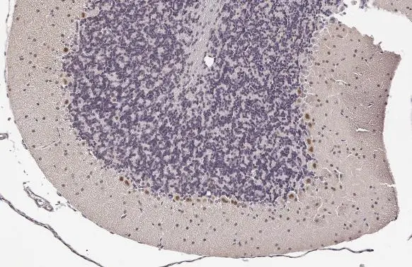
LIM1 antibody detects LIM1 protein at nucleus by immunohistochemical analysis. Sample: Paraffin-embedded mouse cerebellum. LIM1 stained by LIM1 antibody (GTX639639) diluted at 1:100. Antigen Retrieval: Citrate buffer, pH 6.0, 15 min
![LIM1 antibody detects LIM1 protein at nucleus by immunohistochemical analysis. Sample: Paraffin-embedded mouse eye. Green: LIM1 stained by LIM1 antibody (GTX639639) diluted at 1:100. Red: beta Tubulin 3/ Tuj1, a neural marker, stained by beta Tubulin 3/ Tuj1 antibody [GT11710] (GTX631836) diluted at 1:500. Blue: Hoechst 33342 staining. Antigen Retrieval: Citrate buffer, pH 6.0, 15 min](https://www.genetex.com/upload/website/prouct_img/normal/GTX639639/GTX639639_T-45299_20240301_IHC-P_M_1_24030600_629.webp "LIM1 antibody detects LIM1 protein at nucleus by immunohistochemical analysis. Sample: Paraffin-embedded mouse eye. Green: LIM1 stained by LIM1 antibody (GTX639639) diluted at 1:100. Red: beta Tubulin 3/ Tuj1, a neural marker, stained by beta Tubulin 3/ Tuj1 antibody [GT11710] (GTX631836) diluted at 1:500. Blue: Hoechst 33342 staining. Antigen Retrieval: Citrate buffer, pH 6.0, 15 min")
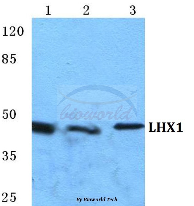
![Various tissue extracts (50 μg) were separated by 10% SDS-PAGE, and the membrane was blotted with LIM1 antibody [HL2774] (GTX639639) diluted at 1:1000. The HRP-conjugated anti-rabbit IgG antibody (GTX213110-01) was used to detect the primary antibody.](https://www.genetex.com/upload/website/prouct_img/normal/GTX639639/GTX639639_45369_20240412_WB_M_tissue_24041523_550.webp "Various tissue extracts (50 μg) were separated by 10% SDS-PAGE, and the membrane was blotted with LIM1 antibody [HL2774] (GTX639639) diluted at 1:1000. The HRP-conjugated anti-rabbit IgG antibody (GTX213110-01) was used to detect the primary antibody.")
LIM1 antibody detects LIM1 protein at nucleus by immunohistochemical analysis. Sample: Paraffin-embedded mouse cerebellum. LIM1 stained by LIM1 antibody (GTX639639) diluted at 1:100. Antigen Retrieval: Citrate buffer, pH 6.0, 15 min
LIM1 antibody [HL2774]
GTX639639
ApplicationsWestern Blot, ImmunoHistoChemistry, ImmunoHistoChemistry Paraffin
Product group Antibodies
ReactivityHuman, Mouse
TargetLHX1
Overview
- SupplierGeneTex
- Product NameLIM1 antibody [HL2774]
- Delivery Days Customer9
- Application Supplier NoteWB: 1:500-1:3000. *Optimal dilutions/concentrations should be determined by the researcher.Not tested in other applications.
- ApplicationsWestern Blot, ImmunoHistoChemistry, ImmunoHistoChemistry Paraffin
- CertificationResearch Use Only
- ClonalityMonoclonal
- Clone IDHL2774
- Concentration1 mg/ml
- ConjugateUnconjugated
- Gene ID3975
- Target nameLHX1
- Target descriptionLIM homeobox 1
- Target synonymsLIM-1, LIM1, LIM/homeobox protein Lhx1, LIM homeobox protein 1, homeobox protein Lim-1
- HostRabbit
- IsotypeIgG
- Protein IDP48742
- Protein NameLIM/homeobox protein Lhx1
- Scientific DescriptionThis gene encodes a member of a large protein family which contains the LIM domain, a unique cysteine-rich zinc-binding domain. The encoded protein is a transcription factor important for the development of the renal and urogenital systems. This gene is a candidate for Mayer-Rokitansky-Kuster-Hauser syndrome, a disorder characterized by anomalies in the female genital tract. [provided by RefSeq, Dec 2010]
- ReactivityHuman, Mouse
- Storage Instruction-20°C or -80°C,2°C to 8°C
- UNSPSC41116161
Datasheet
Related products

Product group Antibodies
Anti-LIM1/LHX1 Antibody Picoband(r)A05816-1-CARRIER-FREE
ApplicationsFlow Cytometry, Western Blot, ELISA, ImmunoHistoChemistry
ReactivityHuman, Mouse
TargetLHX1
- SizePrice
Product group Antibodies
Anti-LHX1 AntibodyA28025
ApplicationsWestern Blot, ImmunoCytoChemistry, ImmunoHistoChemistry
ReactivityHuman, Mouse, Rat
- SizePrice
Product group Antibodies
Anti-LHX1 Antibody144-64893
ApplicationsWestern Blot
ReactivityHuman, Mouse, Rat
TargetLHX1
- SizePrice
Product group Antibodies
LHX1 AntibodyCSB-PA009825
ApplicationsWestern Blot, ELISA, ImmunoHistoChemistry
ReactivityHuman, Mouse, Rat
TargetLHX1
- SizePrice
Product group Antibodies

LHX1 Antibody (Internal)LS-C384339
ApplicationsWestern Blot, ELISA, ImmunoHistoChemistry
ReactivityHuman, Mouse, Rat
TargetLHX1
- SizePrice
Product group Antibodies
LIM1 antibodyGTX13692
ApplicationsWestern Blot
ReactivityHuman
TargetLHX1
- SizePrice
Product group Antibodies
LIM1 antibody, C-termGTX77778
ApplicationsWestern Blot, ImmunoHistoChemistry, ImmunoHistoChemistry Paraffin
ReactivityHuman
TargetLHX1
- SizePrice

Product group Antibodies
Anti-LHX1 AntibodyHPA073521
ApplicationsImmunoHistoChemistry
ReactivityHuman
TargetLHX1
- SizePrice
![LIM1 antibody detects LIM1 protein at cytoplasm and nucleus by immunofluorescent analysis. Sample: DIV9 rat E18 primary cortical neuron cells were fixed in 4% paraformaldehyde at RT for 15 min. Green: LIM1 stained by LIM1 antibody (GTX129215) diluted at 1:250. Red: Tau, an axon marker, stained by Tau antibody [GT287] (GTX634809) diluted at 1:500. Blue: Fluoroshield with DAPI (GTX30920).](https://www.genetex.com/upload/website/prouct_img/normal/GTX129215/GTX129215_44748_20231222_ICC_IF_R_24011618_418.webp)
Product group Antibodies
LIM1 antibodyGTX129215
ApplicationsImmunoFluorescence, ImmunoCytoChemistry
ReactivityHuman, Rat
TargetLHX1
- SizePrice