

Products
Are you looking for life science and diagnostic reagents? We offer one of the most extensive ranges in the Benelux. There are currently more than 5 million products in our webshop, which are manufactured by more than 130 suppliers. This includes both life science and diagnostic reagents. We hope to support your research with everything you need.
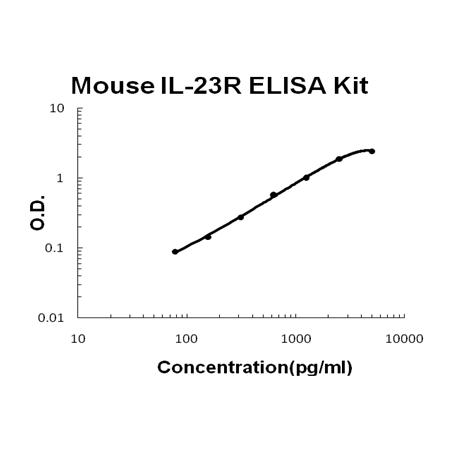
Product group Antibodies
ApplicationsELISA
ReactivityMouse
TargetIl23r
- SizePrice
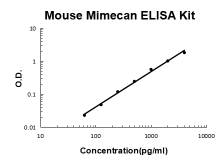
Product group Antibodies
ApplicationsELISA
ReactivityMouse
TargetOgn
- SizePrice
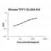
Product group Antibodies
ApplicationsELISA
ReactivityMouse
TargetTff1
- SizePrice
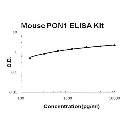
Product group Antibodies
ApplicationsELISA
ReactivityMouse
TargetPon1
- SizePrice
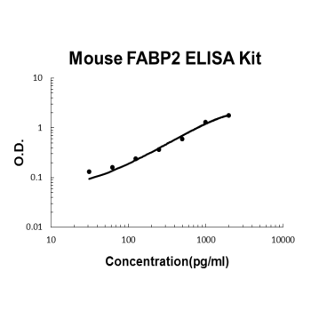
Product group Antibodies
ApplicationsELISA
ReactivityMouse
TargetFabp2
- SizePrice
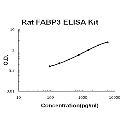
Product group Antibodies
ApplicationsELISA
ReactivityRat
TargetFabp3
- SizePrice
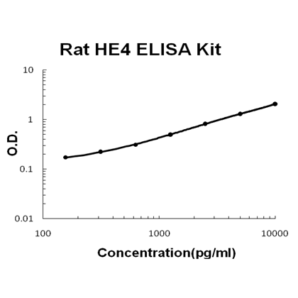
Product group Antibodies
ApplicationsELISA
ReactivityRat
TargetWfdc2
- SizePrice
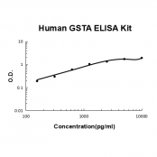
Product group Antibodies
ApplicationsELISA
ReactivityHuman
TargetGSTA1
- SizePrice
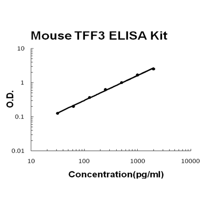
Product group Antibodies
ApplicationsELISA
ReactivityMouse
TargetTff3
- SizePrice
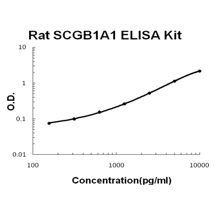
Product group Antibodies
ApplicationsELISA
ReactivityRat
TargetScgb1a1
- SizePrice
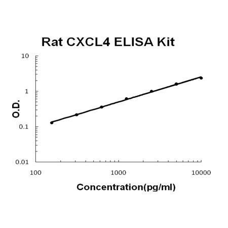
Product group Antibodies
ApplicationsELISA
ReactivityRat
TargetPf4
- SizePrice
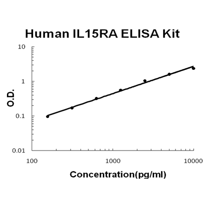
Product group Antibodies
ApplicationsELISA
ReactivityHuman
TargetIL15RA
- SizePrice
Didn't find what you were looking for?
Search through our product groups to find the right product
Back to overview