

Products
Are you looking for life science and diagnostic reagents? We offer one of the most extensive ranges in the Benelux. There are currently more than 5 million products in our webshop, which are manufactured by more than 130 suppliers. This includes both life science and diagnostic reagents. We hope to support your research with everything you need.

Product group Antibodies
ApplicationsELISA
ReactivityHuman
TargetFGF21
- SizePrice
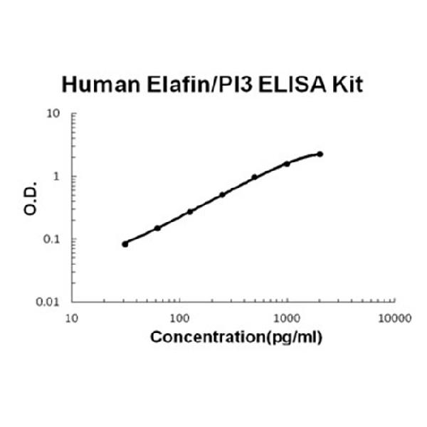
Product group Antibodies
ApplicationsELISA
ReactivityHuman
TargetPI3
- SizePrice
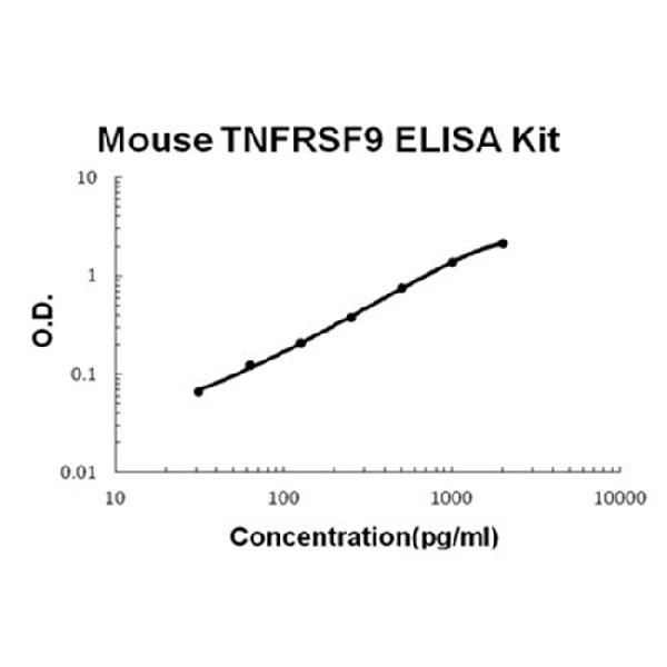
Product group Antibodies
ApplicationsELISA
ReactivityMouse
TargetTnfrsf9
- SizePrice
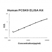
Product group Antibodies
ApplicationsELISA
ReactivityHuman
TargetPCSK9
- SizePrice
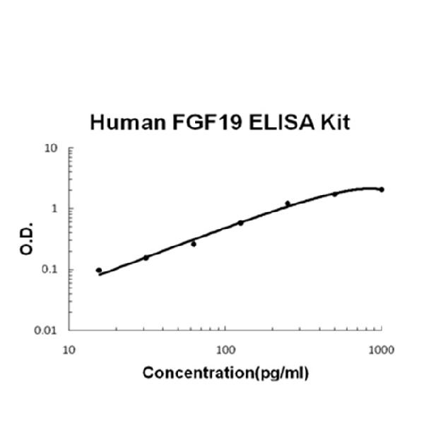
Product group Antibodies
ApplicationsELISA
ReactivityHuman
TargetFGF19
- SizePrice
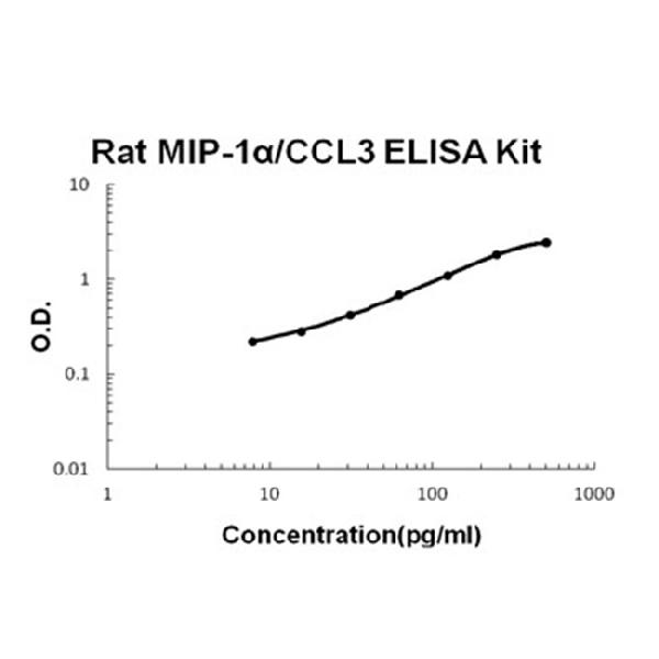
Product group Antibodies
ApplicationsELISA
ReactivityRat
TargetCcl3
- SizePrice
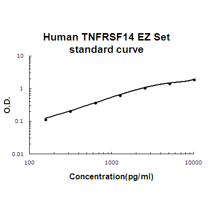
Product group Antibodies
ApplicationsELISA
ReactivityHuman
TargetTNFRSF14
- SizePrice
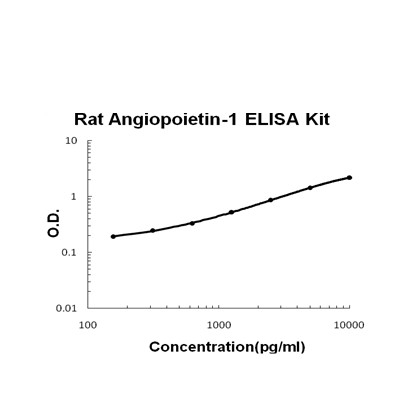
Product group Antibodies
ApplicationsELISA
ReactivityRat
TargetAngpt1
- SizePrice

Product group Antibodies
ApplicationsELISA
ReactivityMouse
TargetAngpt1
- SizePrice
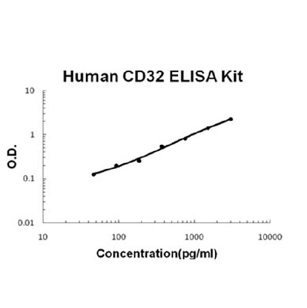
Product group Antibodies
ApplicationsELISA
ReactivityHuman
TargetFCGR2B
- SizePrice

Product group Antibodies
ApplicationsELISA
ReactivityHuman
TargetCRP
- SizePrice
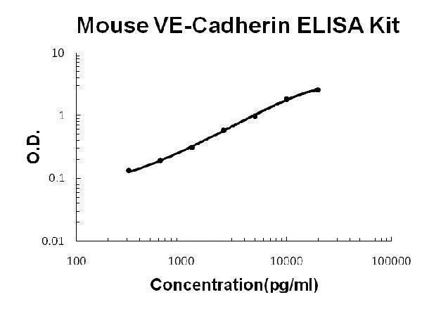
Product group Antibodies
ApplicationsELISA
ReactivityMouse
TargetCdh5
- SizePrice
Didn't find what you were looking for?
Search through our product groups to find the right product
Back to overview