

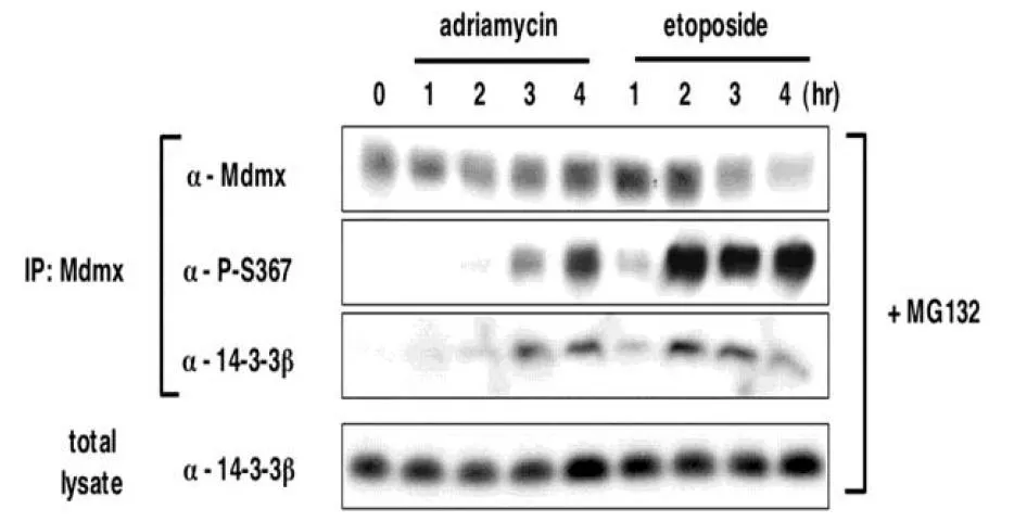
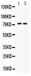
WB analysis of MCF cells with DNA damage using GTX00677 MDMX (phospho Ser367) antibody [#15]. MCF cells were preincubated with the proteasome inhibitor MG132 (20 μM) followed by exposed to DNA damaging agents adriamycin (3 μM) or etoposide (20 μM). The cell lysates were used for immunoprecipitation with anti-MdmX antibody and then analyzed by Western blotting.Induction of S367 phosphorylation after DNA damage is associated with increased binding of 14-3-3 to MdmX and accelerated MdmX degradation.
WB analysis of MCF cells with DNA damage using GTX00677 MDMX (phospho Ser367) antibody [#15]. MCF cells were preincubated with the proteasome inhibitor MG132 (20 μM) followed by exposed to DNA damaging agents adriamycin (3 μM) or etoposide (20 μM). The cell lysates were used for immunoprecipitation with anti-MdmX antibody and then analyzed by Western blotting.Induction of S367 phosphorylation after DNA damage is associated with increased binding of 14-3-3 to MdmX and accelerated MdmX degradation.
MDMX (phospho Ser367) antibody [#15]
GTX00677
ApplicationsImmunoFluorescence, ImmunoPrecipitation, Western Blot, ELISA, ImmunoCytoChemistry
Product group Antibodies
ReactivityHuman, Mouse
TargetMDM4
Overview
- SupplierGeneTex
- Product NameMDMX (phospho Ser367) antibody [#15]
- Delivery Days Customer9
- Application Supplier NoteWB: ~1 microg/ml. *Optimal dilutions/concentrations should be determined by the researcher.Not tested in other applications.
- ApplicationsImmunoFluorescence, ImmunoPrecipitation, Western Blot, ELISA, ImmunoCytoChemistry
- CertificationResearch Use Only
- ClonalityMonoclonal
- Clone ID#15
- Concentration1 mg/ml
- ConjugateUnconjugated
- Gene ID4194
- Target nameMDM4
- Target descriptionMDM4 regulator of p53
- Target synonymsBMFS6, HDMX, MDMX, MRP1, protein Mdm4, MDM4, p53 regulator, MDM4-related protein 1, Mdm4 p53 binding protein homolog, double minute 4, human homolog of; p53-binding protein, mdm2-like p53-binding protein, protein Mdmx
- HostMouse
- IsotypeIgG2b
- Protein IDO15151
- Protein NameProtein Mdm4
- Scientific DescriptionThis gene encodes a nuclear protein that contains a p53 binding domain at the N-terminus and a RING finger domain at the C-terminus, and shows structural similarity to p53-binding protein MDM2. Both proteins bind the p53 tumor suppressor protein and inhibit its activity, and have been shown to be overexpressed in a variety of human cancers. However, unlike MDM2 which degrades p53, this protein inhibits p53 by binding its transcriptional activation domain. This protein also interacts with MDM2 protein via the RING finger domain, and inhibits the latters degradation. So this protein can reverse MDM2-targeted degradation of p53, while maintaining suppression of p53 transactivation and apoptotic functions. Alternatively spliced transcript variants encoding different isoforms have been noted for this gene. [provided by RefSeq, Feb 2011]
- ReactivityHuman, Mouse
- Storage Instruction-20°C or -80°C,2°C to 8°C
- UNSPSC41116161
Datasheet
Related products
Product group Antibodies
Anti-MDM4 AntibodyA96163
ApplicationsWestern Blot, ELISA, ImmunoHistoChemistry
ReactivityHuman, Mouse, Rat
- SizePrice
Product group Antibodies
MDMX Polyclonal AntibodyBS-7663R
ApplicationsImmunoFluorescence, Western Blot, ELISA, ImmunoCytoChemistry, ImmunoHistoChemistry, ImmunoHistoChemistry Frozen, ImmunoHistoChemistry Paraffin
ReactivityBovine, Canine, Equine, Human, Mouse, Porcine, Rabbit, Rat, Sheep
TargetMDM4
- SizePrice

Product group Antibodies
Anti-MDMX/MDM4 Antibody Picoband(r)A01889-2-CARRIER-FREE
ApplicationsWestern Blot, ELISA
ReactivityHuman
TargetMDM4
- SizePrice
Product group Antibodies
MDM4 AntibodyCSB-PA010135
ApplicationsWestern Blot, ELISA, ImmunoHistoChemistry
ReactivityHuman, Mouse, Rat
TargetMDM4
- SizePrice
Product group Antibodies
Mdm4 Polyclonal AntibodyCAC08620
ApplicationsWestern Blot, ELISA
ReactivityMouse
TargetMDM4
- SizePrice
Product group Antibodies

MDM4 / MDMX AntibodyLS-C408345
ApplicationsWestern Blot
ReactivityHuman
TargetMDM4
- SizePrice
Product group Antibodies
MDMX antibodyGTX04638
ApplicationsWestern Blot, ImmunoHistoChemistry, ImmunoHistoChemistry Paraffin
ReactivityHuman, Mouse
TargetMDM4
- SizePrice
Product group Antibodies
MDMX antibody, N-termGTX82948
ApplicationsWestern Blot
ReactivityHuman
TargetMDM4
- SizePrice
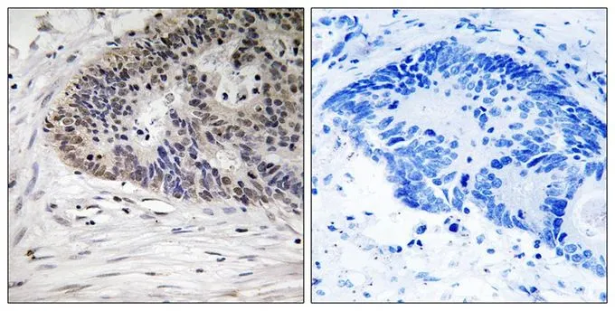
![IHC-P analysis of human cerebra (left) and lung carcinoma (right) tissue using GTX83335 MDMX antibody [2D10F4].](https://www.genetex.com/upload/website/prouct_img/normal/GTX83335/GTX83335_20170912_IHC-P_w_23061322_741.webp)
Product group Antibodies
MDMX antibody [2D10F4]GTX83335
ApplicationsImmunoFluorescence, Western Blot, ELISA, ImmunoCytoChemistry, ImmunoHistoChemistry, ImmunoHistoChemistry Paraffin
ReactivityHuman
TargetMDM4
- SizePrice
Product group Antibodies
MDMX (phospho Ser367) antibodyGTX55445
ApplicationsWestern Blot, ImmunoHistoChemistry, ImmunoHistoChemistry Paraffin
ReactivityHuman
TargetMDM4
- SizePrice