

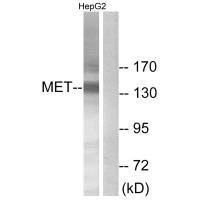
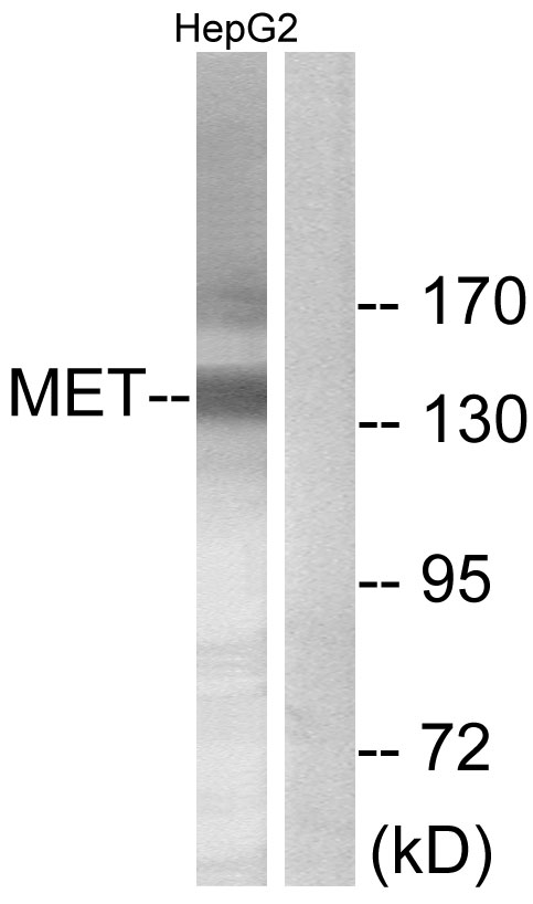
Western blot analysis of extracts from HepG2 cells, using Met (Ab-1234) antibody.
Western blot analysis of extracts from HepG2 cells, using Met (Ab-1234) antibody.
MET (Ab-1234) Antibody
CSB-PA955702
ApplicationsWestern Blot, ELISA
Product group Antibodies
ReactivityHuman, Mouse, Rat
TargetMET
Overview
- SupplierCusabio
- Product NameMET (Ab-1234) Antibody
- Delivery Days Customer20
- ApplicationsWestern Blot, ELISA
- CertificationResearch Use Only
- ClonalityPolyclonal
- ConjugateUnconjugated
- Gene ID4233
- Target nameMET
- Target descriptionMET proto-oncogene, receptor tyrosine kinase
- Target synonymsAUTS9, DA11, DFNB97, HGFR, RCCP2, c-Met, hepatocyte growth factor receptor, HGF receptor, HGF/SF receptor, SF receptor, proto-oncogene c-Met, scatter factor receptor, tyrosine-protein kinase Met
- HostRabbit
- IsotypeIgG
- Protein IDP08581
- Protein NameHepatocyte growth factor receptor
- Scientific DescriptionReceptor tyrosine kinase that transduces signals from the extracellular matrix into the cytoplasm by binding to hepatocyte growth factor/HGF ligand. Regulates many physiological processes including proliferation, scattering, morphogenesis and survival. Ligand binding at the cell surface induces autophosphorylation of MET on its intracellular domain that provides docking sites for downstream signaling molecules. Following activation by ligand, interacts with the PI3-kinase subunit PIK3R1, PLCG1, SRC, GRB2, STAT3 or the adapter GAB1. Recruitment of these downstream effectors by MET leads to the activation of several signaling cascades including the RAS-ERK, PI3 kinase-AKT, or PLCgamma-PKC. The RAS-ERK activation is associated with the morphogenetic effects while PI3K/AKT coordinates prosurvival effects. During embryonic development, MET signaling plays a role in gastrulation, development and migration of muscles and neuronal precursors, angiogenesis and kidney formation. In adults, participates in wound healing as well as organ regeneration and tissue remodeling. Promotes also differentiation and proliferation of hematopoietic cells. Acts as a receptor for Listeria internalin inlB, mediating entry of the pathogen into cells. Gherardi E. et al. (2003).Proc Natl Acad Sci U S A. 100(21): 12039-12044. Shiu SH. et al. (2001) Proc Natl Acad Sci U S A. 98(19): 10763-10768. Hughes AL. et al. (2001) Genome Res. 11(5): 771-780. Onuchic LF. et al. (2002) Am J Hum Genet. 70(5): 1305-13
- ReactivityHuman, Mouse, Rat
- Storage Instruction-20°C or -80°C
- UNSPSC41116161
Related products
Product group Antibodies

Anti-MET [7A2]AB03829-23.0
ApplicationsELISA, Neutralisation/Blocking, Other Application
ReactivityHuman
TargetMET
- SizePrice
Product group Antibodies

c-Met (Phospho-Tyr1003) AntibodyABX012477
ApplicationsImmunoFluorescence, Western Blot, ELISA, ImmunoCytoChemistry, ImmunoHistoChemistry
- SizePrice
![Wild-type (WT) and c-Met knockout (KO) HeLa cell extracts (30 μg) were separated by 5% SDS-PAGE, and the membrane was blotted with c-Met antibody [C3], C-term (GTX100637) diluted at 1:500. The HRP-conjugated anti-rabbit IgG antibody (GTX213110-01) was used to detect the primary antibody.](https://www.genetex.com/upload/website/prouct_img/normal/GTX100637/GTX100637_43901_20200403_WB_KO_watermark_w_23060100_674.webp)
Product group Antibodies
References
c-Met antibody [C3], C-termGTX100637
ApplicationsImmunoFluorescence, ImmunoPrecipitation, Western Blot, ImmunoCytoChemistry
ReactivityHuman
TargetMET
- SizePrice
Product group Antibodies
Anti-METY059019
ApplicationsWestern Blot, ELISA, ImmunoHistoChemistry
ReactivityHuman
- SizePrice
Product group Antibodies
C-Met (MET) Polyclonal AntibodyCAU24571
ApplicationsWestern Blot, ImmunoHistoChemistry
ReactivityMouse
TargetMET
- SizePrice
Product group Antibodies
References
MET Polyclonal AntibodyBS-0668R
ApplicationsFlow Cytometry, ImmunoFluorescence, Western Blot, ELISA, ImmunoCytoChemistry, ImmunoHistoChemistry, ImmunoHistoChemistry Frozen, ImmunoHistoChemistry Paraffin
ReactivityHuman, Mouse, Rat
TargetMET
- SizePrice
Product group Antibodies
Anti-Met AntibodyA101612
ApplicationsWestern Blot, ELISA
ReactivityHuman
- SizePrice