

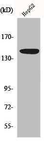
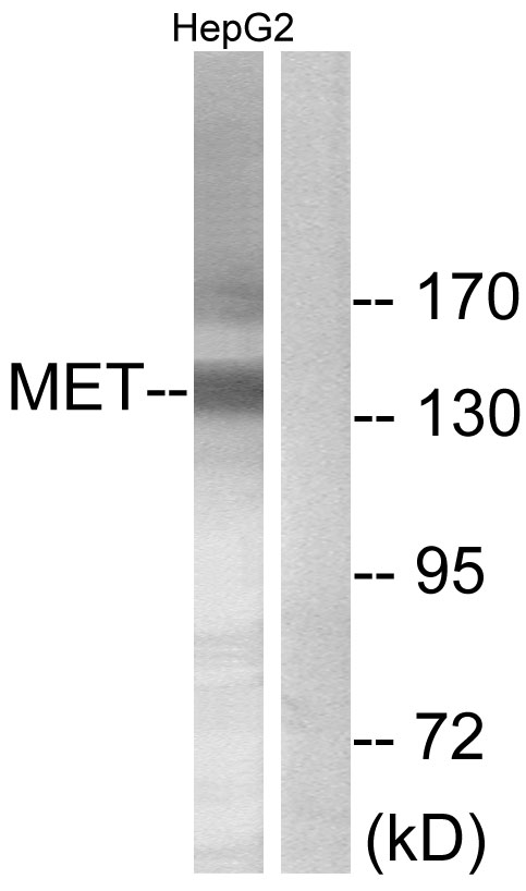
Western Blot analysis of HepG2 cells using Met Polyclonal Antibody
Western Blot analysis of HepG2 cells using Met Polyclonal Antibody
MET Antibody
CSB-PA003231
ApplicationsWestern Blot, ELISA
Product group Antibodies
ReactivityHuman
TargetMET
Overview
- SupplierCusabio
- Product NameMET Antibody
- Delivery Days Customer20
- ApplicationsWestern Blot, ELISA
- CertificationResearch Use Only
- ClonalityPolyclonal
- ConjugateUnconjugated
- Gene ID4233
- Target nameMET
- Target descriptionMET proto-oncogene, receptor tyrosine kinase
- Target synonymsAUTS9, DA11, DFNB97, HGFR, RCCP2, c-Met, hepatocyte growth factor receptor, HGF receptor, HGF/SF receptor, SF receptor, proto-oncogene c-Met, scatter factor receptor, tyrosine-protein kinase Met
- HostRabbit
- IsotypeIgG
- Protein IDP08581
- Protein NameHepatocyte growth factor receptor
- ReactivityHuman
- Storage Instruction-20°C or -80°C
- UNSPSC41116161
Related products
Product group Antibodies

c-Met (Phospho-Tyr1003) AntibodyABX012477
ApplicationsImmunoFluorescence, Western Blot, ELISA, ImmunoCytoChemistry, ImmunoHistoChemistry
- SizePrice
Product group Antibodies
Anti-Met AntibodyA101612
ApplicationsWestern Blot, ELISA
ReactivityHuman
- SizePrice
Product group Antibodies

Anti-MET [7A2]AB03829-23.0
ApplicationsELISA, Neutralisation/Blocking, Other Application
ReactivityHuman
TargetMET
- SizePrice
Product group Antibodies
References
MET Polyclonal AntibodyBS-0668R
ApplicationsImmunoFluorescence, Western Blot, ELISA, ImmunoHistoChemistry, ImmunoHistoChemistry Frozen, ImmunoHistoChemistry Paraffin
ReactivityHuman, Mouse, Rat
TargetMET
- SizePrice
Product group Antibodies
C-Met (MET) Polyclonal AntibodyCAU24571
ApplicationsWestern Blot, ImmunoHistoChemistry
ReactivityMouse
TargetMET
- SizePrice
![Wild-type (WT) and c-Met knockout (KO) HeLa cell extracts (30 μg) were separated by 5% SDS-PAGE, and the membrane was blotted with c-Met antibody [C3], C-term (GTX100637) diluted at 1:500. The HRP-conjugated anti-rabbit IgG antibody (GTX213110-01) was used to detect the primary antibody.](https://www.genetex.com/upload/website/prouct_img/normal/GTX100637/GTX100637_43901_20200403_WB_KO_watermark_w_23060100_674.webp)
Product group Antibodies
c-Met antibody [C3], C-termGTX100637
ApplicationsImmunoFluorescence, ImmunoPrecipitation, Western Blot, ImmunoCytoChemistry
ReactivityHuman
TargetMET
- SizePrice
Product group Antibodies

c-Met Antibody (PE)LS-C486541
ApplicationsFlow Cytometry
ReactivityHuman
TargetMET
- SizePrice

Product group Antibodies
Anti-MET AntibodyHPA055607
ApplicationsImmunoCytoChemistry
ReactivityHuman
TargetMET
- SizePrice