

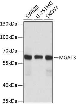
WB analysis of various sample lysates using GTX33317 MGAT3 antibody. Dilution : 1:1000 Loading : 25microg per lane

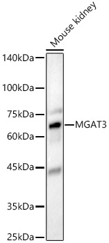
WB analysis of various sample lysates using GTX33317 MGAT3 antibody. Dilution : 1:1000 Loading : 25microg per lane
MGAT3 antibody
GTX33317
ApplicationsWestern Blot, ImmunoHistoChemistry, ImmunoHistoChemistry Paraffin
Product group Antibodies
ReactivityHuman, Rat
TargetMGAT3
Overview
- SupplierGeneTex
- Product NameMGAT3 antibody
- Delivery Days Customer9
- Application Supplier NoteWB: 1:500 - 1:2000. IHC-P: 1:50 - 1:100. *Optimal dilutions/concentrations should be determined by the researcher.Not tested in other applications.
- ApplicationsWestern Blot, ImmunoHistoChemistry, ImmunoHistoChemistry Paraffin
- CertificationResearch Use Only
- ClonalityPolyclonal
- ConjugateUnconjugated
- Gene ID4248
- Target nameMGAT3
- Target descriptionbeta-1,4-mannosyl-glycoprotein 4-beta-N-acetylglucosaminyltransferase
- Target synonymsGNT-III, GNT3, beta-1,4-mannosyl-glycoprotein 4-beta-N-acetylglucosaminyltransferase, GlcNAc-T III, N-acetylglucosaminyltransferase III, N-glycosyl-oligosaccharide-glycoprotein N-acetylglucosaminyltransferase III, mannosyl (beta-1,4-)-glycoprotein beta-1,4-N-acetylglucosaminyltransferase
- HostRabbit
- IsotypeIgG
- Protein IDQ09327
- Protein NameBeta-1,4-mannosyl-glycoprotein 4-beta-N-acetylglucosaminyltransferase
- Scientific DescriptionThere are believed to be over 100 different glycosyltransferases involved in the synthesis of protein-bound and lipid-bound oligosaccharides. The enzyme encoded by this gene transfers a GlcNAc residue to the beta-linked mannose of the trimannosyl core of N-linked oligosaccharides and produces a bisecting GlcNAc. Multiple alternatively spliced variants, encoding the same protein, have been identified. [provided by RefSeq, Jul 2008]
- ReactivityHuman, Rat
- Storage Instruction-20°C or -80°C,2°C to 8°C
- UNSPSC12352203
Datasheet
Related products
Product group Antibodies
Mgat3 Polyclonal AntibodyCAC11386
ApplicationsWestern Blot, ELISA, ImmunoHistoChemistry
ReactivityMouse
TargetMGAT3
- SizePrice
Product group Antibodies
Anti-MGAT3 AntibodyA16082
ApplicationsWestern Blot
ReactivityHuman, Mouse, Rat
- SizePrice
Product group Antibodies
Anti-MGAT3 Antibody144-08134
ApplicationsWestern Blot, ImmunoHistoChemistry
ReactivityHuman, Mouse, Rat
TargetMGAT3
- SizePrice
Product group Antibodies
References
MGAT3 antibody [N3C3]GTX112153
ApplicationsImmunoFluorescence, Western Blot, ImmunoCytoChemistry, ImmunoHistoChemistry, ImmunoHistoChemistry Paraffin
ReactivityHuman, Mouse
TargetMGAT3
- SizePrice
Product group Antibodies

GNT-III / MGAT3 AntibodyLS-C409670
ApplicationsWestern Blot, ImmunoHistoChemistry
ReactivityHuman, Mouse, Rat
TargetMGAT3
- SizePrice

Product group Antibodies
Anti-MGAT3 AntibodyHPA017598
ApplicationsImmunoHistoChemistry
ReactivityHuman
TargetMGAT3
- SizePrice
Product group Antibodies
MGAT3 AntibodyCSB-PA22969A0RB
ApplicationsWestern Blot, ELISA, ImmunoHistoChemistry
ReactivityHuman, Mouse
TargetMGAT3
- SizePrice

Product group Antibodies
Anti-MGAT3 Antibody Picoband(r)A08470-1-CARRIER-FREE
ApplicationsWestern Blot, ELISA, ImmunoHistoChemistry
ReactivityHuman
TargetMGAT3
- SizePrice