

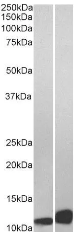
Various whole cell extracts (30 μg) were separated by 15% SDS-PAGE, and the membrane was blotted with MIF antibody [HL2963] (GTX640350) diluted at 1:1000. The HRP-conjugated anti-rabbit IgG antibody (GTX213110-01) was used to detect the primary antibody.
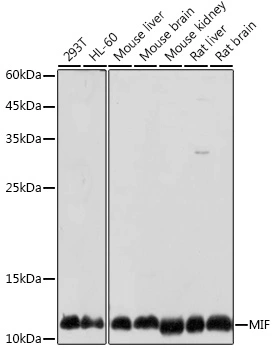
![Various tissue extracts (50 μg) were separated by 15% SDS-PAGE, and the membrane was blotted with MIF antibody [HL2963] (GTX640350) diluted at 1:1000. The HRP-conjugated anti-rabbit IgG antibody (GTX213110-01) was used to detect the primary antibody.](https://www.genetex.com/upload/website/prouct_img/normal/GTX640350/GTX640350_45481_20240729_WB_M_tissue_24080602_499.webp "Various tissue extracts (50 μg) were separated by 15% SDS-PAGE, and the membrane was blotted with MIF antibody [HL2963] (GTX640350) diluted at 1:1000. The HRP-conjugated anti-rabbit IgG antibody (GTX213110-01) was used to detect the primary antibody.")
![Indirect ELISA analysis was performed by coating the plate with recombinant Mammalian cell expressed, full-length human MIF protein (96.85-1.51 nM). Coated protein was probed with MIF antibody [HL2963] (GTX640350) (1 μg/mL). Goat anti-rabbit IgG antibody (HRP) (GTX213110-01) (1:10000) was used to detect the bound primary antibody.](https://www.genetex.com/upload/website/prouct_img/normal/GTX640350/GTX640350_T-45411_20240906_ELISA_Indirect_24091102_533.webp "Indirect ELISA analysis was performed by coating the plate with recombinant Mammalian cell expressed, full-length human MIF protein (96.85-1.51 nM). Coated protein was probed with MIF antibody [HL2963] (GTX640350) (1 μg/mL). Goat anti-rabbit IgG antibody (HRP) (GTX213110-01) (1:10000) was used to detect the bound primary antibody.")
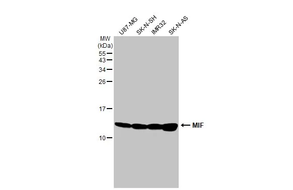
Various whole cell extracts (30 μg) were separated by 15% SDS-PAGE, and the membrane was blotted with MIF antibody [HL2963] (GTX640350) diluted at 1:1000. The HRP-conjugated anti-rabbit IgG antibody (GTX213110-01) was used to detect the primary antibody.
MIF antibody [HL2963]
GTX640350
ApplicationsWestern Blot, ELISA
Product group Antibodies
ReactivityHuman, Mouse
TargetMIF
Overview
- SupplierGeneTex
- Product NameMIF antibody [HL2963]
- Delivery Days Customer7
- Application Supplier NoteWB: 1:500-1:3000. *Optimal dilutions/concentrations should be determined by the researcher.Not tested in other applications.
- ApplicationsWestern Blot, ELISA
- CertificationResearch Use Only
- ClonalityMonoclonal
- Clone IDHL2963
- Concentration1 mg/ml
- ConjugateUnconjugated
- Gene ID4282
- Target nameMIF
- Target descriptionmacrophage migration inhibitory factor
- Target synonymsGIF, GLIF, MMIF, macrophage migration inhibitory factor, L-dopachrome isomerase, L-dopachrome tautomerase, epididymis secretory sperm binding protein, macrophage migration inhibitory factor (glycosylation-inhibiting factor), phenylpyruvate tautomerase
- HostRabbit
- IsotypeIgG
- Protein IDP14174
- Protein NameMacrophage migration inhibitory factor
- Scientific DescriptionThis gene encodes a lymphokine involved in cell-mediated immunity, immunoregulation, and inflammation. It plays a role in the regulation of macrophage function in host defense through the suppression of anti-inflammatory effects of glucocorticoids. This lymphokine and the JAB1 protein form a complex in the cytosol near the peripheral plasma membrane, which may indicate an additional role in integrin signaling pathways. [provided by RefSeq, Jul 2008]
- ReactivityHuman, Mouse
- Storage Instruction-20°C or -80°C,2°C to 8°C
- UNSPSC12352203
Datasheet
Related products
Product group Antibodies
Anti-MIF Antibody130-10009
ApplicationsWestern Blot, ELISA
ReactivityHuman
- SizePrice
Product group Antibodies

Kininogen 1 (KNG1) AntibodyABX109959
ApplicationsImmunoFluorescence, Western Blot, ELISA, ImmunoCytoChemistry, ImmunoHistoChemistry
- SizePrice
Product group Antibodies
Mif Polyclonal AntibodyCAC07247
ApplicationsImmunoFluorescence, Western Blot, ELISA, ImmunoHistoChemistry
ReactivityMouse, Rat
TargetMIF
- SizePrice
Product group Antibodies
References
MIF Polyclonal AntibodyBS-1044R
ApplicationsFlow Cytometry, ImmunoFluorescence, Western Blot, ELISA, ImmunoCytoChemistry, ImmunoHistoChemistry, ImmunoHistoChemistry Frozen, ImmunoHistoChemistry Paraffin
TargetMIF
- SizePrice
Product group Antibodies
ApplicationsWestern Blot, ELISA
ReactivityHuman
- SizePrice
Product group Antibodies
MIF antibody, C-termGTX89535
ApplicationsWestern Blot, ELISA, ImmunoHistoChemistry, ImmunoHistoChemistry Paraffin
ReactivityHuman
TargetMIF
- SizePrice
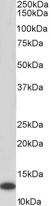
![Western blot analysis of GeneTex's IgY fraction of Chicken-anti-Human MIF polyclonal antibody shows the detection of 100 μg of recombinant MIF present in a lysate. Similar detection of MIF will occur when human serum is analyzed. In lane 1 no reaction is observed in the control whereas lane 2 shows a single band at 12.3 kDa. A 4-20% gradient gel was used to separate the proteins by SDS-PAGE. The protein was transferred to nitro-cellulose using standard methods. After blocking the membrane was probed with the primary antibody for 1 h at room temperature followed by washes and reaction with a 1:5,000 dilution of IRDye800 conjugated Gt-a-Chicken Rabbit IgG [H&L] for 1 h at room temperature. LICOR's OdysseyR Infrared Imaging System was used to scan and process the image. Other detection systems will yield similar results.](https://www.genetex.com/upload/website/prouct_img/normal/GTX48478/GTX48478_20160330_WB_w_23060823_846.webp)
Product group Antibodies
MIF antibodyGTX48478
ApplicationsWestern Blot, ELISA
ReactivityHuman
TargetMIF
- SizePrice
Product group Antibodies
MIF ELISA PairGTX500014
ApplicationsELISA
ReactivityHuman, Mouse
TargetMIF
- SizePrice
Product group Antibodies
MIF antibodyGTX55704
ApplicationsWestern Blot
ReactivityHuman, Mouse, Rat
TargetMIF
- SizePrice