

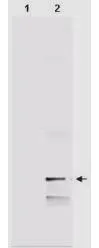
Western blot analysis of GeneTex's IgY fraction of Chicken-anti-Human MIF polyclonal antibody shows the detection of 100 μg of recombinant MIF present in a lysate. Similar detection of MIF will occur when human serum is analyzed. In lane 1 no reaction is observed in the control whereas lane 2 shows a single band at 12.3 kDa. A 4-20% gradient gel was used to separate the proteins by SDS-PAGE. The protein was transferred to nitro-cellulose using standard methods. After blocking the membrane was probed with the primary antibody for 1 h at room temperature followed by washes and reaction with a 1:5,000 dilution of IRDye800 conjugated Gt-a-Chicken Rabbit IgG [H&L] for 1 h at room temperature. LICOR's OdysseyR Infrared Imaging System was used to scan and process the image. Other detection systems will yield similar results.

Western blot analysis of GeneTex's IgY fraction of Chicken-anti-Human MIF polyclonal antibody shows the detection of 100 μg of recombinant MIF present in a lysate. Similar detection of MIF will occur when human serum is analyzed. In lane 1 no reaction is observed in the control whereas lane 2 shows a single band at 12.3 kDa. A 4-20% gradient gel was used to separate the proteins by SDS-PAGE. The protein was transferred to nitro-cellulose using standard methods. After blocking the membrane was probed with the primary antibody for 1 h at room temperature followed by washes and reaction with a 1:5,000 dilution of IRDye800 conjugated Gt-a-Chicken Rabbit IgG [H&L] for 1 h at room temperature. LICOR's OdysseyR Infrared Imaging System was used to scan and process the image. Other detection systems will yield similar results.
MIF antibody
GTX48478
ApplicationsWestern Blot, ELISA
Product group Antibodies
ReactivityHuman
TargetMIF
Overview
- SupplierGeneTex
- Product NameMIF antibody
- Delivery Days Customer9
- Application Supplier NoteWB: 1:500-1:2000. ELISA: 1:1000-1:5000. *Optimal dilutions/concentrations should be determined by the researcher.Not tested in other applications.
- ApplicationsWestern Blot, ELISA
- CertificationResearch Use Only
- ClonalityPolyclonal
- Concentration5 mg/ml
- ConjugateUnconjugated
- Gene ID4282
- Target nameMIF
- Target descriptionmacrophage migration inhibitory factor
- Target synonymsGIF, GLIF, MMIF, macrophage migration inhibitory factor, L-dopachrome isomerase, L-dopachrome tautomerase, epididymis secretory sperm binding protein, macrophage migration inhibitory factor (glycosylation-inhibiting factor), phenylpyruvate tautomerase
- HostChicken
- IsotypeIgY
- Protein IDP14174
- Protein NameMacrophage migration inhibitory factor
- Scientific DescriptionPro-inflammatory cytokine. Involved in the innate immune response to bacterial pathogens. The expression of MIF at sites of inflammation suggests a role as mediator in regulating the function of macrophages in host defense. Counteracts the anti-inflammatory activity of glucocorticoids. Has phenylpyruvate tautomerase and dopachrome tautomerase activity (in vitro), but the physiological substrate is not known. It is not clear whether the tautomerase activity has any physiological relevance, and whether it is important for cytokine activity.
- ReactivityHuman
- Storage Instruction-20°C or -80°C,2°C to 8°C
- UNSPSC41116161
Datasheet
Related products
Product group Antibodies
MIF AntibodyCSB-PA003240
ApplicationsWestern Blot, ELISA
ReactivityHuman, Mouse, Rat
TargetMIF
- SizePrice
Product group Antibodies
ApplicationsWestern Blot, ELISA
ReactivityHuman
- SizePrice
Product group Antibodies

Kininogen 1 (KNG1) AntibodyABX109959
ApplicationsImmunoFluorescence, Western Blot, ELISA, ImmunoCytoChemistry, ImmunoHistoChemistry
- SizePrice
Product group Antibodies

MIF Antibody (Preservative Free)LS-C149173
ApplicationsWestern Blot, ELISA
ReactivityHuman
TargetMIF
- SizePrice
Product group Antibodies

Goat anti-MIF, BiotinylatedEB06765-B
ApplicationsWestern Blot, ELISA
ReactivityHuman
TargetMIF
- SizePrice

Product group Antibodies
Anti-MIF AntibodyHPA003868
ApplicationsWestern Blot, ImmunoCytoChemistry, ImmunoHistoChemistry
ReactivityHuman, Mouse, Rat
TargetMIF
- SizePrice
Product group Antibodies
MIF antibody [2Ar3]GTX14575
ApplicationsWestern Blot
ReactivityHuman
TargetMIF
- SizePrice
Product group Antibodies
Mif Polyclonal AntibodyCAC07247
ApplicationsImmunoFluorescence, Western Blot, ELISA, ImmunoHistoChemistry
ReactivityMouse, Rat
TargetMIF
- SizePrice