

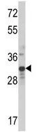
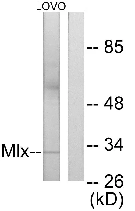
WB analysis of MDA-MB231 cell lysate (35ug/lane) using GTX81125 MLX antibody, Internal.
 either nontransfected (Lane 1) or transiently transfected with the MLX (Lane 2) using GTX81125 MLX antibody, Internal.")


WB analysis of MDA-MB231 cell lysate (35ug/lane) using GTX81125 MLX antibody, Internal.
MLX antibody, Internal
GTX81125
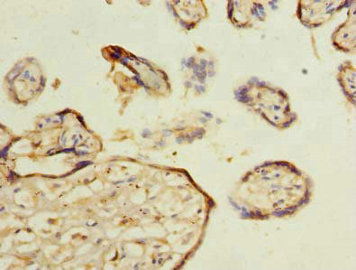
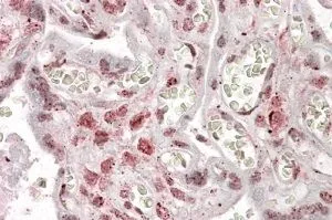
ApplicationsFlow Cytometry, Western Blot, ImmunoHistoChemistry, ImmunoHistoChemistry Paraffin
Product group Antibodies
ReactivityHuman
TargetMLX
Overview
- SupplierGeneTex
- Product NameMLX antibody, Internal
- Delivery Days Customer9
- Application Supplier NoteWB: 1:1000. IHC-P: 1:50-1:100. FCM: 1:10-1:50. *Optimal dilutions/concentrations should be determined by the researcher.Not tested in other applications.
- ApplicationsFlow Cytometry, Western Blot, ImmunoHistoChemistry, ImmunoHistoChemistry Paraffin
- CertificationResearch Use Only
- ClonalityPolyclonal
- ConjugateUnconjugated
- Gene ID6945
- Target nameMLX
- Target descriptionMAX dimerization protein MLX
- Target synonymsMAD7, MXD7, TCFL4, TF4, bHLHd13, max-like protein X, BigMax protein, MAX-like bHLHZIP protein, MLX, MAX dimerization protein, class D basic helix-loop-helix protein 13, transcription factor-like protein 4
- HostRabbit
- IsotypeIgG
- Protein IDQ9UH92
- Protein NameMax-like protein X
- Scientific DescriptionThe product of this gene belongs to the family of basic helix-loop-helix leucine zipper (bHLH-Zip) transcription factors. These factors form heterodimers with Mad proteins and play a role in proliferation, determination and differentiation. This gene product may act to diversify Mad family function by its restricted association with a subset of the Mad family of transcriptional repressors, namely, Mad1 and Mad4. Alternatively spliced transcript variants encoding different isoforms have been identified for this gene. [provided by RefSeq, Jul 2008]
- ReactivityHuman
- Storage Instruction-20°C or -80°C,2°C to 8°C
- UNSPSC41116161
Datasheet
Related products
Product group Antibodies
MLX AntibodyCSB-PA014643LA01HU
ApplicationsImmunoFluorescence, ELISA, ImmunoHistoChemistry
ReactivityHuman
TargetMLX
- SizePrice

Product group Antibodies
Anti-MLX Antibody Picoband(r)A03087-2-CARRIER-FREE
ApplicationsFlow Cytometry, Western Blot
ReactivityHuman, Monkey, Mouse
TargetMLX
- SizePrice

Product group Antibodies
Anti-MLX AntibodyHPA052766
ApplicationsImmunoHistoChemistry
ReactivityHuman
TargetMLX
- SizePrice
Product group Antibodies

Goat anti-MLXEB09756
ApplicationsELISA, ImmunoHistoChemistry
ReactivityBovine, Canine, Human, Mouse, Rat
TargetMLX
- SizePrice
Product group Antibodies

MLX / TCFL4 Antibody (HRP)LS-C376128
ApplicationsELISA, ImmunoHistoChemistry
ReactivityHuman
TargetMLX
- SizePrice
Product group Antibodies
MLX antibodyGTX87667
ApplicationsWestern Blot
ReactivityHuman
TargetMLX
- SizePrice
Product group Antibodies
MLX antibody, InternalGTX88235
ApplicationsImmunoHistoChemistry, ImmunoHistoChemistry Paraffin
ReactivityHuman
TargetMLX
- SizePrice
Product group Antibodies
MLX Polyclonal AntibodyBS-3950R
ApplicationsImmunoFluorescence, Western Blot, ELISA, ImmunoCytoChemistry, ImmunoHistoChemistry, ImmunoHistoChemistry Frozen, ImmunoHistoChemistry Paraffin
ReactivityBovine, Canine, Chicken, Equine, Human, Mouse, Porcine, Rabbit, Rat
TargetMLX
- SizePrice