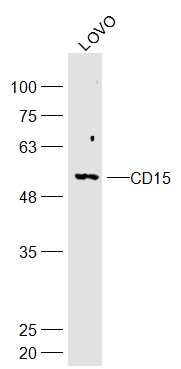
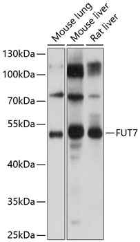
Mouse anti Human CD15
0151
ApplicationsFlow Cytometry
Product group Antibodies
TargetFUT4
Overview
- SupplierNordic-MUbio
- Product NameMouse anti Human CD15
- Delivery Days Customer7
- Application Supplier NotePBMC: Add10 microl of MAB/10^6 PBMC in 100 microl PBS. Mix gently and incubate for 15 minutes at 2 to 8C. Wash twice with PBS and analyze or fix with 0.5% v/v of paraformaldehyde in PBS and analyze. WHOLE BLOOD: Add 10 microl of MAB/100 microl of whole blood. Mix gently and incubate for 15 minutes at room temperature (20C). Lyse the whole blood. Wash once with PBS and analyze or fix with 0.5% v/v of paraformaldehyde in PBS and analyze. See instrument manufacturers instructions for Lysed Whole Blood and Immunofluorescence analysis with a flow cytometer or microscope.
- ApplicationsFlow Cytometry
- Applications SupplierFlow Cytometry
- CertificationResearch Use Only
- ClonalityMonoclonal
- Clone IDARE
- ConjugateUnconjugated
- Gene ID2526
- Target nameFUT4
- Target descriptionfucosyltransferase 4
- Target synonymsCD15, ELFT, FCT3A, FUC-TIV, FUTIV, LeX, SSEA-1, alpha-(1,3)-fucosyltransferase 4, 4-galactosyl-N-acetylglucosaminide 3-alpha-L-fucosyltransferase, ELAM ligand fucosyltransferase, ELAM-1 ligand fucosyltransferase, Lewis X, alpha (1,3) fucosyltransferase, myeloid-specific, fucT-IV, fucosyltransferase IV, galactoside 3-L-fucosyltransferase, stage-specific embryonic antigen 1
- HostMouse
- IsotypeIgM
- Protein IDP22083
- Protein NameAlpha-(1,3)-fucosyltransferase 4
- Scientific DescriptionCD15
- Shelf life instructionSee expiration date on vial
- Reactivity SupplierHuman
- UNSPSC12352203


