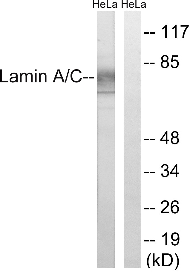
Mouse anti Lamin A and C, conjugated to FITC
MUB1102L1
ApplicationsFlow Cytometry, ImmunoCytoChemistry, ImmunoHistoChemistry, ImmunoHistoChemistry Frozen
Product group Antibodies
ReactivityBovine, Canine, Hamster, Human, Mouse, Rat, Sheep
TargetLMNA
Overview
- SupplierNordic-MUbio
- Product NameMouse anti Lamin A and C, conjugated to FITC
- Delivery Days Customer7
- Application Supplier Note131C3 is suitable for immunocytochemistry, immunohistochemistry on frozen sections and flow cytometry. Optimal antibody dilutions for the different applications should be determined by titration. The recommended dilution is 1:10.
- ApplicationsFlow Cytometry, ImmunoCytoChemistry, ImmunoHistoChemistry, ImmunoHistoChemistry Frozen
- Applications SupplierFlow Cytometry;Immunocytochemistry;Immunohistochemistry (frozen)
- CertificationResearch Use Only
- ClonalityMonoclonal
- Clone ID131C3
- ConjugateFITC
- Gene ID4000
- Target nameLMNA
- Target descriptionlamin A/C
- Target synonymsCDCD1, CDDC, CMD1A, CMT2B1, EMD2, FPL, FPLD, FPLD2, HGPS, IDC, LDP1, LFP, LGMD1B, LMN1, LMNC, LMNL1, MADA, PRO1, lamin, 70 kDa lamin, epididymis secretory sperm binding protein, lamin A/C-like 1, lamin C, mandibuloacral dysplasia type A, prelamin-A/C, progerin, renal carcinoma antigen NY-REN-32
- HostMouse
- IsotypeIgG1
- Protein IDP02545
- Protein NamePrelamin-A/C
- Source131C3 is a mouse monoclonal IgG1/k antibody derived by fusion of P3/X63.Ag8.653 mouse myeloma cells with spleen cells from a BALB/c mouse immunized with purified rat liver lamins.
- ReactivityBovine, Canine, Hamster, Human, Mouse, Rat, Sheep
- Reactivity SupplierBovine;Dog;Hamster;Human;Mouse;Rat;Sheep
- UNSPSC12352203


