

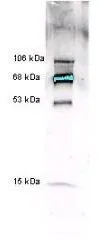
Anti-Myeloperoxidase [Human Leukocytes] detects multiple MPO subunits and chain combinations by western blot. Polyclonal rabbit-anti-Myeloperoxidase (GTX22088) was used at a 1:5000 dilution to detect 1.0 ug of human myeloperoxidase. This antibody detects a multiple bands corresponding to 53 kDa and 15 kDa polypeptides and chain combinations forming 68 kDa and 106 kDa proteins. The staining of the 68 kDa band is so intense that is over saturates the signal detection. A 4-20% gradient gel was used to separate the protein by SDS-PAGE. The protein was transferred to nitrocellulose using standard methods. After blocking the membrane was probed with the primary antibody for 2 h at room temperature followed by washes and reaction with a 1:5,000 dilution of IRDye?800 conjugated Goat anti Rabbit IgG [H&L] for 30 min at room temperature.

Anti-Myeloperoxidase [Human Leukocytes] detects multiple MPO subunits and chain combinations by western blot. Polyclonal rabbit-anti-Myeloperoxidase (GTX22088) was used at a 1:5000 dilution to detect 1.0 ug of human myeloperoxidase. This antibody detects a multiple bands corresponding to 53 kDa and 15 kDa polypeptides and chain combinations forming 68 kDa and 106 kDa proteins. The staining of the 68 kDa band is so intense that is over saturates the signal detection. A 4-20% gradient gel was used to separate the protein by SDS-PAGE. The protein was transferred to nitrocellulose using standard methods. After blocking the membrane was probed with the primary antibody for 2 h at room temperature followed by washes and reaction with a 1:5,000 dilution of IRDye?800 conjugated Goat anti Rabbit IgG [H&L] for 30 min at room temperature.
Myeloperoxidase antibody
GTX22088
ApplicationsImmunoPrecipitation, Western Blot, ELISA
Product group Antibodies
TargetMPO
Overview
- SupplierGeneTex
- Product NameMyeloperoxidase antibody
- Delivery Days Customer9
- Application Supplier NoteWB: 1:5000. IP: 1:100. ELISA: 1:200000. *Optimal dilutions/concentrations should be determined by the researcher.Not tested in other applications.
- ApplicationsImmunoPrecipitation, Western Blot, ELISA
- CertificationResearch Use Only
- ClonalityPolyclonal
- Concentration75 mg/ml
- ConjugateUnconjugated
- Gene ID4353
- Target nameMPO
- Target descriptionmyeloperoxidase
- Target synonymsmyeloperoxidase
- HostRabbit
- IsotypeIgG
- Protein IDP05164
- Protein NameMyeloperoxidase
- Scientific DescriptionHuman myeloperoxidase (MPO) is a dimeric protein composed of two heavy subunits (53 kDa) and two light subunits (15 kDa). Each MPO molecule contains two prosthetic porphyrins which play an important role in the catalytic cycle. Molecular weights for MPO isoforms from pools of normal human samples range from 114,000 to 140,000 daltons reflecting a heterogeneous mixture of isoforms when assayed under non-reducing conditions of SDS-PAGE. Often MPO from a single donor will yield a homogenous preparation reflecting a single isoform. The carbohydrate component of MPO, consisting of mannose, glucose and N-acetylglucosamine residues is 2.5%. MPO is inhibited by azide and other compounds. MPO is stored in primary granules of neutrophils and serves as a bactericidal agent in that MPO catalyzes the production of hypochlorous acid (HOCl), a powerful oxidant. HOCl is derived from chloride ion (Cl-) and hydrogen peroxide (H2O2). In a number of inflammatory situations, MPO is released into the extracellular matrix where its measurement can be used as an indication of neutrophil activation.
- Storage Instruction-20°C or -80°C,2°C to 8°C
- UNSPSC12352203
References
- The first reported case of trastuzumab induced interstitial lung disease associated with anti-neutrophil cytoplasmic antibody vasculitis - A case report and a prospective cohort study on the usefulness of neutrophil derived biomarkers in monitoring vasculitis disease activity during follow-up. Chang CH et al., 2022 Feb, BreastRead more
Datasheet
Related products
Product group Antibodies
MPO AntibodyCSB-PA005559
ApplicationsWestern Blot, ELISA
ReactivityHuman, Mouse, Rat
TargetMPO
- SizePrice
Product group Antibodies
Myeloperoxidase antibodyGTX135125
ApplicationsImmunoFluorescence, Western Blot, ImmunoCytoChemistry
TargetMPO
- SizePrice
Product group Antibodies
Myeloperoxidase antibodyGTX135126
ApplicationsImmunoFluorescence, Western Blot, ImmunoCytoChemistry, ImmunoHistoChemistry, ImmunoHistoChemistry Paraffin
TargetMPO
- SizePrice
Product group Antibodies
Myeloperoxidase antibodyGTX135127
ApplicationsImmunoFluorescence, Western Blot, ImmunoCytoChemistry
TargetMPO
- SizePrice
Product group Antibodies

Mouse anti Myeloperoxidase (MPO)GM-4191-CE/IVD
ApplicationsFlow Cytometry
TargetMPO
- SizePrice
![IHC-P analysis of human bone marrow, granulocytic sarcoma using GTX01896 Myeloperoxidase antibody [59A5]. Note intense cytoplasmic staining of malignant myeloid cells.](https://www.genetex.com/upload/website/prouct_img/normal/GTX01896/GTX01896_20200811_IHC-P_99_w_23053121_318.webp)
Product group Antibodies
Myeloperoxidase antibody [59A5]GTX01896
ApplicationsImmunoHistoChemistry, ImmunoHistoChemistry Paraffin
TargetMPO
- SizePrice
Product group Antibodies
References
Myeloperoxidase antibody [2C7]GTX75315
ApplicationsFlow Cytometry, Western Blot, ELISA, ImmunoHistoChemistry, ImmunoHistoChemistry Frozen, ImmunoHistoChemistry Paraffin
TargetMPO
- SizePrice
Product group Antibodies
References
Myeloperoxidase antibody [2C7]GTX75318
ApplicationsFlow Cytometry, ImmunoFluorescence, Western Blot, ELISA, ImmunoCytoChemistry, ImmunoHistoChemistry, ImmunoHistoChemistry Frozen, ImmunoHistoChemistry Paraffin
TargetMPO
- SizePrice
Product group Antibodies
Myeloperoxidase antibody [16E3]GTX82940
ApplicationsELISA, Purification/Extraction/Isolation
TargetMPO
- SizePrice