

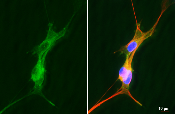
Neurokinin 1 Receptor antibody [HL3194] detects Neurokinin 1 Receptor protein by immunofluorescent analysis. Sample: U87-MG cells were fixed in 4% paraformaldehyde at RT for 15 min. Green: Neurokinin 1 Receptor stained by Neurokinin 1 Receptor antibody [HL3194] (GTX640833) diluted at 1:500. Red: alpha Tubulin, a cytoskeleton marker, stained by alpha Tubulin antibody [GT114] (GTX628802) diluted at 1:1000. Blue: Fluoroshield with DAPI (GTX30920).
![Neurokinin 1 Receptor antibody [HL3194] detects Neurokinin 1 Receptor protein by immunofluorescent analysis. Sample: Mock and transfected 293T cells were fixed in ice-cold MeOH for 5 min. Green: Neurokinin 1 Receptor stained by Neurokinin 1 Receptor antibody [HL3194] (GTX640833) diluted at 1:500. Blue: Fluoroshield with DAPI (GTX30920).](https://www.genetex.com/upload/website/prouct_img/normal/GTX640833/GTX640833_T-45495_20240906_ICC_IF_B_24092600_260.webp "Neurokinin 1 Receptor antibody [HL3194] detects Neurokinin 1 Receptor protein by immunofluorescent analysis. Sample: Mock and transfected 293T cells were fixed in ice-cold MeOH for 5 min. Green: Neurokinin 1 Receptor stained by Neurokinin 1 Receptor antibody [HL3194] (GTX640833) diluted at 1:500. Blue: Fluoroshield with DAPI (GTX30920).")
Neurokinin 1 Receptor antibody [HL3194] detects Neurokinin 1 Receptor protein by immunofluorescent analysis. Sample: U87-MG cells were fixed in 4% paraformaldehyde at RT for 15 min. Green: Neurokinin 1 Receptor stained by Neurokinin 1 Receptor antibody [HL3194] (GTX640833) diluted at 1:500. Red: alpha Tubulin, a cytoskeleton marker, stained by alpha Tubulin antibody [GT114] (GTX628802) diluted at 1:1000. Blue: Fluoroshield with DAPI (GTX30920).
Neurokinin 1 Receptor antibody [HL3194]
GTX640833
ApplicationsImmunoFluorescence, ImmunoCytoChemistry
Product group Antibodies
ReactivityHuman
TargetTACR1
Overview
- SupplierGeneTex
- Product NameNeurokinin 1 Receptor antibody [HL3194]
- Delivery Days Customer7
- Application Supplier NoteICC/IF: 1:100-1:1000. *Optimal dilutions/concentrations should be determined by the researcher.Not tested in other applications.
- ApplicationsImmunoFluorescence, ImmunoCytoChemistry
- CertificationResearch Use Only
- ClonalityMonoclonal
- Clone IDHL3194
- Concentration1 mg/ml
- ConjugateUnconjugated
- Gene ID6869
- Target nameTACR1
- Target descriptiontachykinin receptor 1
- Target synonymsNK1R, NKIR, SPR, TAC1R, substance-P receptor, NK-1 receptor, NK-1R, neurokinin receptor 1, tachykinin receptor 1 (substance P receptor; neurokinin-1 receptor)
- HostRabbit
- IsotypeIgG
- Protein IDP25103
- Protein NameSubstance-P receptor
- Scientific DescriptionThis gene belongs to a gene family of tachykinin receptors. These tachykinin receptors are characterized by interactions with G proteins and contain seven hydrophobic transmembrane regions. This gene encodes the receptor for the tachykinin substance P, also referred to as neurokinin 1. The encoded protein is also involved in the mediation of phosphatidylinositol metabolism of substance P. [provided by RefSeq, Sep 2008]
- ReactivityHuman
- Storage Instruction-20°C or -80°C,2°C to 8°C
- UNSPSC41116161
Datasheet
Related products
Product group Antibodies
Anti-TACR1 AntibodyA37627
ApplicationsWestern Blot, ImmunoHistoChemistry
ReactivityHuman, Mouse, Rat
- SizePrice
Product group Antibodies
Anti-TACR1 (C-term) Antibody102-24500
ApplicationsWestern Blot
TargetTACR1
- SizePrice
Product group Antibodies
References
ApplicationsWestern Blot, ELISA
ReactivityBovine, Canine, Equine, Guinea Pig, Human, Mouse, Porcine, Rat
TargetTACR1
- SizePrice
Product group Antibodies

Goat anti-TACR1EB07771
ApplicationsImmunoFluorescence, Western Blot, ELISA
ReactivityCanine, Human, Mouse
TargetTACR1
- SizePrice
Product group Antibodies
TACR1 AntibodyCSB-PA050201
ApplicationsImmunoFluorescence, Western Blot, ELISA
ReactivityHuman, Mouse, Rat
TargetTACR1
- SizePrice
Product group Antibodies
ApplicationsImmunoPrecipitation, Western Blot, ImmunoCytoChemistry, ImmunoHistoChemistry
TargetTACR1
- SizePrice
Product group Antibodies
Neurokinin 1 Receptor antibodyGTX30183
ApplicationsFlow Cytometry, Western Blot, ImmunoHistoChemistry, ImmunoHistoChemistry Paraffin
ReactivityGuinea Pig, Human, Monkey, Mouse, Rat
TargetTACR1
- SizePrice

Product group Antibodies
Anti-TACR1 AntibodyHPA074573
ApplicationsImmunoHistoChemistry
ReactivityHuman
TargetTACR1
- SizePrice
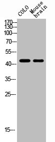
![Rat tissue extract (50 μg) was separated by 10% SDS-PAGE, and the membrane was blotted with Neurokinin 1 Receptor antibody [HL3195] (GTX640834) diluted at 1:1000. The HRP-conjugated anti-rabbit IgG antibody (GTX213110-01) was used to detect the primary antibody.](https://www.genetex.com/upload/website/prouct_img/normal/GTX640834/GTX640834_T-45495_20240809_WB_R_brain_24081300_888.webp)
Product group Antibodies
ApplicationsWestern Blot, ImmunoHistoChemistry, ImmunoHistoChemistry Paraffin
ReactivityCanine, Human, Mouse, Rat
TargetTACR1
- SizePrice