

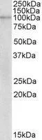
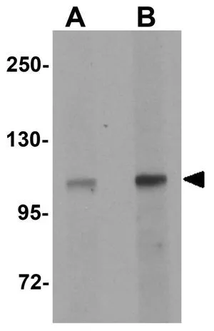
WB analysis of human breast cancer lysate using GTX88038 NLRX1 antibody, Internal. Dilution : 2μg/ml Loading : 35μg protein in RIPA buffer
WB analysis of human breast cancer lysate using GTX88038 NLRX1 antibody, Internal. Dilution : 2μg/ml Loading : 35μg protein in RIPA buffer
NLRX1 antibody, Internal
GTX88038
ApplicationsWestern Blot
Product group Antibodies
ReactivityHuman
TargetNLRX1
Overview
- SupplierGeneTex
- Product NameNLRX1 antibody, Internal
- Delivery Days Customer7
- Application Supplier NoteWB: 1-3microg/ml. *Optimal dilutions/concentrations should be determined by the researcher.Not tested in other applications.
- ApplicationsWestern Blot
- CertificationResearch Use Only
- ClonalityPolyclonal
- Concentration0.50 mg/ml
- ConjugateUnconjugated
- Gene ID79671
- Target nameNLRX1
- Target descriptionNLR family member X1
- Target synonymsCLR11.3, DLNB26, NOD26, NOD5, NOD9, NLR family member X1, NOD-like receptor X1, caterpiller protein 11.3, nucleotide-binding oligomerization domain protein 26, nucleotide-binding oligomerization domain protein 5, nucleotide-binding oligomerization domain protein 9, nucleotide-binding oligomerization domain, leucine rich repeat containing X1
- HostGoat
- IsotypeIgG
- Protein IDQ86UT6
- Protein NameNLR family member X1
- Scientific DescriptionThe protein encoded by this gene is a member of the NLR family and localizes to the outer mitochondrial membrane. The encoded protein is a regulator of mitochondrial antivirus responses. Three transcript variants encoding the same protein have been found for this gene. [provided by RefSeq, Aug 2013]
- ReactivityHuman
- Storage Instruction-20°C or -80°C,2°C to 8°C
- UNSPSC12352203
Datasheet
Related products
Product group Antibodies
Anti-NLRX1 Antibody144-61405
ApplicationsWestern Blot
ReactivityHuman, Mouse, Rat
TargetNLRX1
- SizePrice
Product group Antibodies
NLRX1 Polyclonal AntibodyBS-55154R
ApplicationsWestern Blot
ReactivityHuman, Mouse, Rat
TargetNLRX1
- SizePrice
Product group Antibodies
Anti-NLRX1 AntibodyA95824
ApplicationsWestern Blot, ELISA, ImmunoHistoChemistry
ReactivityHuman, Mouse, Rat
- SizePrice
Product group Antibodies
NLRX1 AntibodyCSB-PA015878LA01HU
ApplicationsELISA, ImmunoHistoChemistry
ReactivityHuman
TargetNLRX1
- SizePrice

Product group Antibodies
Anti-NLRX1 AntibodyHPA061516
ApplicationsImmunoCytoChemistry, ImmunoHistoChemistry
ReactivityHuman
TargetNLRX1
- SizePrice
Product group Antibodies

ApplicationsWestern Blot, ELISA
ReactivityHuman, Rat
TargetNLRX1
- SizePrice
Product group Antibodies

NLRX1 AntibodyLS-C333745
ApplicationsWestern Blot
ReactivityHuman, Mouse, Rat
TargetNLRX1
- SizePrice
Product group Antibodies
NLRX1 antibodyGTX84999
ApplicationsWestern Blot, ELISA, ImmunoHistoChemistry, ImmunoHistoChemistry Paraffin
ReactivityHuman, Mouse, Rat
TargetNLRX1
- SizePrice

Product group Antibodies
Anti-NLRX1 Antibody Picoband(r)A04980-3-CARRIER-FREE
ApplicationsFlow Cytometry, ImmunoFluorescence, Western Blot, ELISA, ImmunoCytoChemistry, ImmunoHistoChemistry
ReactivityHuman, Mouse, Rat
TargetNLRX1
- SizePrice