

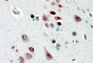
IHC-P analysis of human cerebral cortex using GTX88815 NOVA1 antibody, Internal. Antigen retrieval : citrate buffer pH 6 Dilution : 2.5μg/ml
 using GTX88815 NOVA1 antibody, Internal.")

IHC-P analysis of human cerebral cortex using GTX88815 NOVA1 antibody, Internal. Antigen retrieval : citrate buffer pH 6 Dilution : 2.5μg/ml
NOVA1 antibody, Internal
GTX88815
ApplicationsWestern Blot, ImmunoHistoChemistry, ImmunoHistoChemistry Paraffin
Product group Antibodies
ReactivityHuman
TargetNOVA1
Overview
- SupplierGeneTex
- Product NameNOVA1 antibody, Internal
- Delivery Days Customer9
- Application Supplier NoteWB: 0.01-0.03microg/ml. IHC-P: 2-4microg/ml. *Optimal dilutions/concentrations should be determined by the researcher.Not tested in other applications.
- ApplicationsWestern Blot, ImmunoHistoChemistry, ImmunoHistoChemistry Paraffin
- CertificationResearch Use Only
- ClonalityPolyclonal
- Concentration0.50 mg/ml
- ConjugateUnconjugated
- Gene ID4857
- Target nameNOVA1
- Target descriptionNOVA alternative splicing regulator 1
- Target synonymsNova-1, RNA-binding protein Nova-1, neuro-oncological ventral antigen 1, onconeural ventral antigen 1, paraneoplastic Ri antigen, ventral neuron-specific protein 1
- HostGoat
- IsotypeIgG
- Protein IDP51513
- Protein NameRNA-binding protein Nova-1
- Scientific DescriptionThis gene encodes a neuron-specific RNA-binding protein, a member of the Nova family of paraneoplastic disease antigens, that is recognized and inhibited by paraneoplastic antibodies. These antibodies are found in the sera of patients with paraneoplastic opsoclonus-ataxia, breast cancer, and small cell lung cancer. Alternatively spliced transcripts encoding distinct isoforms have been described. [provided by RefSeq, Jul 2008]
- ReactivityHuman
- Storage Instruction-20°C or -80°C,2°C to 8°C
- UNSPSC41116161
Datasheet
Related products
Product group Antibodies
NOVA1 AntibodyCSB-PA015957LA01HU
ApplicationsWestern Blot, ELISA, ImmunoHistoChemistry
ReactivityHuman
TargetNOVA1
- SizePrice

Product group Antibodies
Anti-Nova1 Antibody Picoband(r)A06087-1-CARRIER-FREE
ApplicationsFlow Cytometry, ImmunoFluorescence, Western Blot, ELISA, ImmunoCytoChemistry, ImmunoHistoChemistry, ImmunoHistoChemistry Frozen
ReactivityHuman, Mouse, Rat
TargetNOVA1
- SizePrice
Product group Antibodies

NOVA1 AntibodyLS-C747238
ApplicationsWestern Blot
ReactivityHuman
TargetNOVA1
- SizePrice
Product group Antibodies

Goat anti-NOVA1EB08387
ApplicationsWestern Blot, ELISA, ImmunoHistoChemistry
ReactivityHuman
TargetNOVA1
- SizePrice

Product group Antibodies
Anti-NOVA1 AntibodyHPA004155
ApplicationsWestern Blot, ImmunoCytoChemistry, ImmunoHistoChemistry
ReactivityHuman
TargetNOVA1
- SizePrice
Product group Antibodies
NOVA1 Polyclonal AntibodyCAC13137
ApplicationsWestern Blot, ELISA, ImmunoHistoChemistry
TargetNOVA1
- SizePrice
Product group Antibodies
NOVA1 antibodyGTX102831
ApplicationsImmunoFluorescence, Western Blot, ImmunoCytoChemistry
ReactivityHuman
TargetNOVA1
- SizePrice
Product group Antibodies
Anti-NOVA1 (Center) Antibody102-24148
ApplicationsWestern Blot, ImmunoHistoChemistry, ImmunoHistoChemistry Paraffin
TargetNOVA1
- SizePrice